|
Fig. 1 |
Fig. 2 |
Fig. 3 |
|
ESPAÑOL |
ENGLISH |
|
CASO CLÍNICO 13 Autores: JV Bagán (*), E Lloria (*), F Cardona (*), MA Milián (*), Y Jiménez (*), A Pérez (*) (*)Servicio de Estomatología del Hospital General Universitario de Valencia. Medicina Bucal de la Universidad de Valencia. |
CLINICAL CASE 13 Authors: JV Bagán (*), E Lloria (*), F Cardona (*), MA Milián (*), Y Jiménez (*), A Pérez (*) (*)Servicio de Estomatología del Hospital General Universitario de Valencia. Medicina Bucal de la Universidad de Valencia. |
|
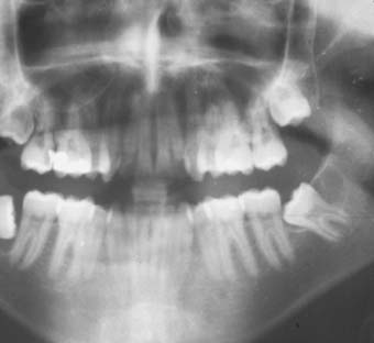
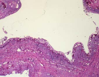
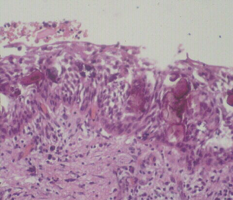
SÍNTOMAS Y EXPLORACIÓN CLÍNICA Paciente de 33 años que tenía los terceros molares inferiores impactados. No tenía síntomas. La ortopantomografía demostraba una imagen quística alrededor de la corona del izquierdo (38) (Fig. 1) . El molar fue extraído y el quiste enviado para biopsia (Fig. 2 y 3) |
SYMPTOMS AND CLINICAL EXAMINATION A 33 year-old man presented with impacted third mandibular molars. He hadn´t symtoms. The ortopanthomograph showed a cystic cavity around the crown of the left third molar (38) (Fig. 1) . The molar was extracted and the cystic sac was submitted for biopsy (Fig. 2 y 3) |
|
Fig. 1 |
Fig. 2 |
Fig. 3 |
|
DIAGNÓSTICOS PROPUESTOS |
PROVISIONAL DIAGNOSIS |
|
Quiste odontogénico calcificante Quiste folicular Quiste paradental |
Calcifying odontogenic cyst Dentigerous cyst Paradental cyst |
|
DEFINITIVE DIAGNOSIS |
|
|
Quiste odontogénico calcificante |
Calcifying odontogenic cyst |
CASOS
/ CASES© José V. Bagán . Universidad de Valencia (España)