|
|
|
|
(*) Servicio de Estomatología. Hospital General Universitario de Valencia. Medicina Bucal, Universidad de Valencia. (Spain) |
(*) Servicio de Estomatología. Hospital General Universitario de Valencia. Medicina Bucal, Universidad de Valencia. (Spain) |
|
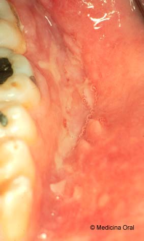
Varón de 23 años que
presenta desde hace un mes unas lesiones erosivas en su mucosa oral. No
ha tomado fármacos ni había tenido estas lesiones en otras
ocasiones.
A la exploración intraoral
se aprecian lesiones erosivas grandes, en algunos casos de más de
3 cm. de largo, mal definidas y con un carácter ligeramente granular
y duro en algunos puntos. Son especialmente evidentes en paladar y
fondos de vestíbulos tanto inferiores como inferiores (Figura
1) (Figura 2).
|
A 23-year-old male presented a one-month history of erosive lesions affecting the oral mucosa. He was not taking medication and had not suffered such lesions in the past. The lesions were painful and made eating difficult. In addition, the patient suffered digestive disorders with diarrhea, for several months. Intraoral examination revealed large, poorly defined erosive lesions (in some cases over 3 cm in length); the latter were slightly granular and hard at certain points, and were particularly evident on the palate and in both the lower and upper vestibular cavities (Figure 1) . (Figure 2). A biopsy was performed of the lower vestibular fundus (Figure 3) (Figura 4) .
|
|
Enfermedad de Crohn Tuberculosis Sarcoidosis Micosis profunda Colitis ulcerosa |
Crohn disease |
|
|
|
|
|
|
|
|
Agradecimiento: A Alvaro Carpi por el procesado de las muestras
histológicas.