|
CASO CLÍNICO 8
Autores:
Francisco Cardona, José V.
Bagán, M. Angeles Milián, Enrique Lloria, Yolanda Jiménez.
(*) Servicio de Estomatología.
Hospital General Universitario de Valencia. Medicina Bucal, Universidad
de Valencia. (Spain) |
CLINICAL CASE 8
Authors:
Francisco Cardona, José V.
Bagán, M. Angeles Milián, Enrique Lloria, Yolanda Jiménez.
(*) Servicio de Estomatología.
Hospital General Universitario de Valencia. Medicina Bucal, Universidad
de Valencia. (Spain) |
SÍNTOMAS Y EXPLORACIÓN
CLÍNICA
Anamnesis y exploración física
Varón de 41 años que presenta un crecimiento
progresivo e indoloro del maxilar superior en la zona de premolares del
lado derecho, desde hace unos pocos años (Figura
1). La lesión crea una cierta deformidad facial, aunque no
molestias. No existen otras lesiones óseas.
Exploraciones complementarias
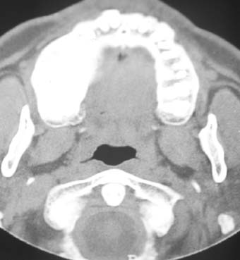
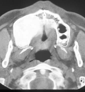
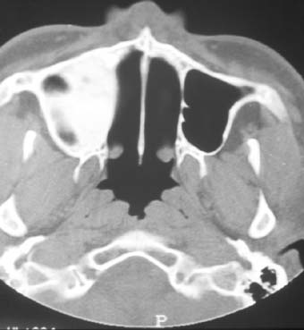
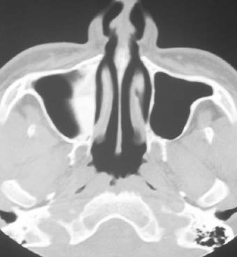
La ortopantomografía y la TAC mostraba una extensa
área radiopaca en el maxilar superior
(Figura 2)(Figura 3) (Figura 4)(Figura
5)
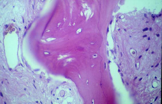
Se tomó una biopsia del hueso maxilar
(Figura 6). Los análisis de sangre fueron normales, incluso
las fosfatas alcalinas. |
SYMPTOMS AND CLINICAL EXAMINATION
Anamnesis and physical exploration
A 41-year-old male presented a progressive painless growth
in the upper maxilla at right premolar level, for the past few years (Figure
1). The lesion caused facial deformity, though no physical discomfort.
No bony lesions were detected elsewhere.
Complementary explorations
Orthopantomography and CAT revealed an extensive radiopaque
zone in the upper maxilla (Figure 2) (Figure 3) (Figure
4)(Figure 5)
The maxillary bone was biopsied (Figure
6). Blood tests were normal, including alkaline phosphatase levels. |