MEDICINA ORAL  

CASOS CLINICOS / CLINICAL CASES

Colaboración científica / Scientific collaboration:
Prof. Heddie O. Sedano (Los Angeles, USA) 

INFORMACIÓN (Español) / INFORMATION (English)


ESPAÑOL
ENGLISH
CASO CLÍNICO 9 
Autores:  

José V. Bagán,  Miguel Peñarrocha, M. Ángeles Milián, Yolanda Jiménez. 
(*) Servicio de Estomatología. Hospital General Universitario de Valencia. Medicina Bucal, Universidad de Valencia. (Spain) 

CLINICAL CASE 9  
Authors:  

José V. Bagán,  Miguel Peñarrocha, M. Ángeles Milián, Yolanda Jiménez. 
(*) Servicio de Estomatología. Hospital General Universitario de Valencia. Medicina Bucal, Universidad de Valencia. (Spain) 

SÍNTOMAS Y EXPLORACIÓN CLÍNICA 
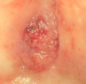
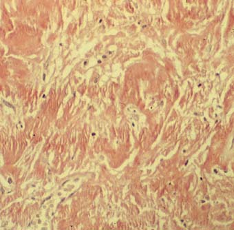
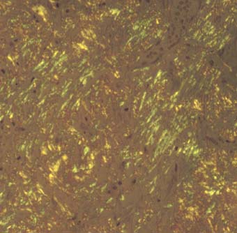
Paciente de 73 años que desde hacía 3 meses presentaba una lesión tumoral en paladar que le originaba hemorragias espontáneas, habiendo crecido de tamaño poco a poco hasta la situación actual. No le producía dolor ni molestias. No había ningún foco dental ni óseo que lo pudiese justificar. 
A la exploración intraoral se apreciaba una tumoración de 1.5 x 1.5 cm en el centro del paladar, de color rojo, bien delimitada (figura 1). En la ortopantomografía y TAC no se evidenciaba lesión en hueso maxilar, siendo la lesión exclusiva de mucosa. 
Se tomó una biopsia de la lesión (figura 2) (figura 3). 
Los resultados de pruebas de coagulación fueron normales, no tenía lesiones cutáneas, ni digestivas, tampoco alteraciones renales, cardiacas, hepáticas ni neurológicas. En la analítica hemática y de orina no había alteraciones ni proteinas anómalas. 
 
SYMPTOMS AND CLINICAL EXAMINATION 

Male 73 years old with a 3 month history of a palatal mass. The tumor which had a steady 
growth, bled spontaneously and it was non painful. There were neither dental nor bony lesions 
associated with it. 
Clinical intraoral exam revealed a well delineated red mass in the center of the palate measuring 1.5 by 1.5 cm.  (figure 1) 
Orthopantomography and CAT did not revealed any bony lesion. 
Review of system did not show any abnormalities in neither, skin, GI tract, kidney, myocardium, liver and CNS. Coagulation time as well as blood an urine analyses were within normal limits. 
A biopsy of the lesion was undertaken. (figure 2) (figure 3) 


figure 1
figure 2 (H-E)
figure 3 (Congo red)
Pulse cualquiera de las imágenes para verlas en tamaño ampliado
Press in the figures to a higher magnification
 
 
DIAGNÓSTICOS PROPUESTOS 
PROVISIONAL DIAGNOSIS 
-Amiloidosis primaria
-Primary amyloidosis
 
 
DIAGNÓSTICO DEFINITIVO 
DEFINITIVE DIAGNOSIS 
Amiloidosis primaria
Primary amyloidosis


CASOS / CASES
 
Listado de casos y diagnósticos
Cases and diagnosis

 
 © José V. Bagán . Universidad de Valencia (España) e-mail: bagan@uv.es