|
Gorka Bastarrika G y col. New techniques for the evaluation
and therapeutic planning of patients with KlippeleTre´naunay syndrome J Am Acad Dermatol 2007;56:242-9.) |
|
|
| Fig 1. A, Geographic stain on thigh and
knee of a 26-year-old female with KlippeleTre´naunay syndrome (patient 5). B, MDCT images. C, Volume rendered images of the right lower extremity. Severely dilated lateral veins (arrows) extending into the pelvis. Antero-posterior view. D, Duplication of the superficial femoral vein (arrows). Note a normal superficial femoral vein in the contralateral limb (arrowhead ). Posterio-anterior view. |
|
|
| Fig 2. A, Geographic stain on lateral thigh,
knee, and leg of a 34-year-old male with KlippeleTre´naunay syndrome (patient 7). B, MR venography. Persistent lower sciatic veins (arrow) originate in the region of the popliteal vein and terminate in the proximal superficial femoral vein (arrowhead ). |
  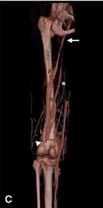 |
| Fig 3. A, Geographic stain on the foot of a
24-year-old male with KlippeleTre´naunay syndrome (patient 8). B, MDCT images. C, MDCT venography. Normal superficial femoral vein (arrow) with aplasia of the popliteal vein (arrowhead ), and a persistent sciatic vein (asterisk). |


