- by Dr. Rafael Sebastian Aguilar
- 2015
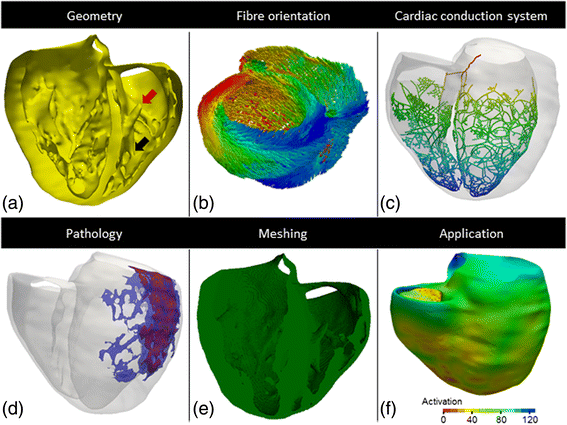
Electrophysiology Model:
3 Cell Types (Endo-, Mid-, Epi-)Anatomy Model:
Includes trabeculas and Fiber orientationReference Paper:
10.1186/s12938-015-0033-5Download:
Ventricular Volume Hex Model (VTK)Ventricular Surface Tris Model (VTK)
Ventricular Purkinje Model (VTK)
Models review table (PDF)
- by Dr. Rafael Sebastian Aguilar
- 2019
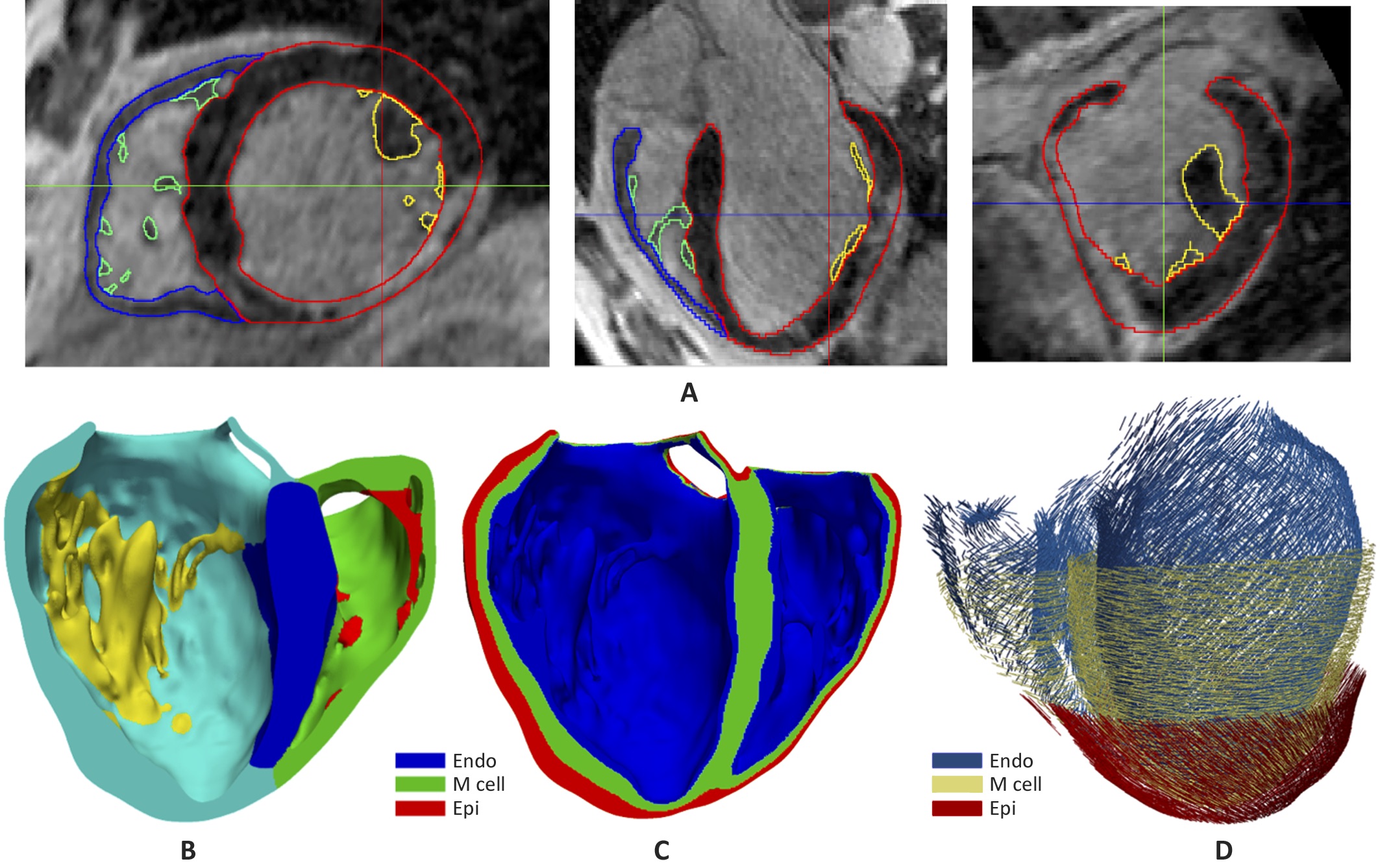
Electrophysiology Model:
3 Cell Types (Endo-, Mid-, Epi-)Fitted endocardial CARTO maps
Anatomical Model and Scar:
The model was segmented with endocardial trabeculas. Model includes segmentation of the scar with border zone and core zone. It is embedded in a 3D torso model including, bones, lungs, liver, and atria. Endocardial and epicardial Electro-anatomical maps (EAM) of the patient fit to the model are included.Reference Paper:
10.3389/fphys.2019.00580Simulations during ventricular tachycardia:
YouTube VideoDownload:
Ventricular Volume Model + Labels + Scar (Hexahedra VTK)Ventricular Surface Model + Labels + Scar (3 x VTK files)Ventricular Purkinje Stochastic Model (VTK)
Electro-anatomical data fit to model -CARTO Measure Points- (3 x VTK files)
Electro-anatomical data fit to model -CARTO Surfaces- (3 x VTK files)Full Labelled Torso Model (VTK)
- by Pau Romero De Antonio, Dr. Ignacio Garcia-Fernandez and Dr. Miguel Lozano Ibañez
- 2021
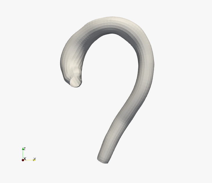
Anatomy Model:
3000 thoracic aortas. Include sinuses of Valsalva.No supra-aortic branches.
Regular triangle meshes with 150 (longitudinal) x 40 (transversal) subdivisions.
Reference Paper:
10.3389/fphys.2021.713118Download:
STL Files

- by Dr. Miguel Rodrigo Bort
- 2023
Simulation of atrial fibrillation sustained by a microanatomic reentry and the simultaneous EGM registered with a unipolar electrode at 2mm and 6mm from the pectinate muscle, respectively.
- by Dr. Rafael Sebastian Aguilar
- 2015
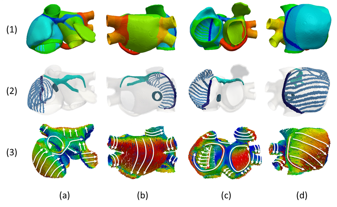
We have developed a new atrial model that improves our previous 3D model of the human atria. The new model (available online at Download Media section) has enhanced anatomical and functional heterogeneity and the detailed regional description of fibre direction. The resulting computational finite element model is a multi-layer mesh with a homogeneous wall thickness between 600 and 900 μm (754.893 nodes and 515.010 elements), built with linear hexahedral elements with regular spatial resolution of 300 µm. Following a histological analysis, the model was manually divided into 21 regions. However, for the detailed description of fibre orientation, some regions were further subdivided into a total of 53 sub-regions. The model also includes the preferential conduction bundles (Fig. 2 row 2), which are the CT, PMs, BB with right (BBR) and left (BBL) sides and the LFO between both atria. Atrial fibre orientation was analysed in detail to determine the number and location of regions with a common fibre organization (Fig. 2 row 3). This orientation was assigned to each region as the cross product between the principal region-defining vector direction and the normal to the external surface of each element belonging to that region.
Electrophysiology Models:
8 Cell Types, 7 Tissue TypesAnatomy Model:
53 Regions Fiber Orientation directions, Voxel: 0.3 mmReference Paper:
10.1371/journal.pone.0141573Download:
3D Atria Model (VTK)Cellular parameters (TXT)
Full Details (PDF)

- by Dr. Rafael Sebastian Aguilar
- 2015