MEDICINA ORAL


CASOS CLÍNICOS / CLINICAL CASES

Editores/Editors:
Prof. Jose V. Bagan (University of Valencia - Spain)
Prof. Crispian Scully (University of London - U.K.) 

Colaboración científica / Scientific collaboration:
Prof. Heddie O. Sedano (Los Angeles, USA) 

INFORMACIÓN (Español) / INFORMATION (English)


ESPAÑOL
ENGLISH
CASO CLÍNICO 6 
Autor: Jose M. Aguirre
Medicina Bucal, Universidad del País Vasco. (Spain)
CLINICAL CASE 6 
Author: Jose M. Aguirre
Oral Medicine. País Vasco University (Spain)
SÍNTOMAS Y EXPLORACIÓN CLÍNICA
 

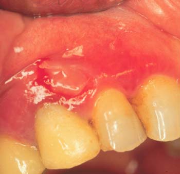
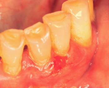
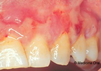
Mujer de 67 años que presenta desde hace un año y medio unas lesiones en encías que le producen dolor y escozor. A la exploración oral se observan áreas eritematosas en encías de ambos maxilares, tanto en la vertiente vestibular como lingual, acompañándose de zonas ampollares sangrantes y descamativas  (Figura 1) (Figura 2) . El signo de Nikolsky fue negativo. No existían alteraciones en otras mucosas ni en la piel. 
Se tomaron muestras para estudio histopatológico  (Figura 3) (Figura 4) e inmunofluorescencia directa en la que manifestó una positividad con la IgA en zona de membrana basal   (Figura 5). 

SYMPTOMS AND CLINICAL EXAMINATION
 
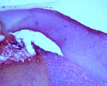
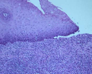
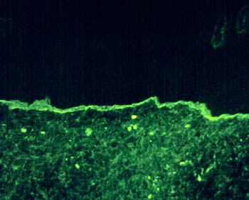
A 67 year old female with an 18 months history of various painful and burning gingival lesions. Intraoral exam revealed 
erythematous lesions on the vestibular and lingual, maxillary and mandibular gingiva  (Figure 1) (Figure 2) . A vesiculo-erosive eruption was also noted. The vesicles and bullae had a bleeding tendency. The Nikolsky sign was negative. They were no other mucosal or skin lesions. Biopsies were taken for routine histopathologic  (Figure 3) (Figure 4) and immunofluorescence  in which we found and IgA positivity at basement membrane zone (Figure 5)
DIAGNÓSTICOS PROVISIONALES PROPUESTOS POR AQUELLOS QUE ENVIARON UN E-mail 
Pengigoide benigno de las mucosas
Penpigo
Liquen plano
PROVISIONAL DIAGNOSIS PROPOSED FOR PARTICIPANTS (Those who sent an E-mail)
Mucous membrane penphigoid
Penphigus
Lichen planus 


COMENTARIOS
Debemos señalar que inicialmente no se señaló cuando presentamos el caso clínico que la inmunofluorescencia era positiva para la IgA, simplemente se puso una imagen con una positividad en zona de membrana basal. 


In the first presentation we didn´t say the positivity at basement membrane zone was with IgA


DIAGNÓSTICO DEFINITIVO
 
DEFINITIVE DIAGNOSIS
 
Dermatosis IgA linear de la infancia
Linear IgA disease

© 1996-1997 Medicina Oral
Prof. José V. Bagan
UNIVERSIDAD DE VALENCIA (ESPAÑA)
VALENCIA UNIVERSITY (SPAIN)