MEDICINA ORAL


CASOS CLÍNICOS / CLINICAL CASES

Editores/Editors:
Prof. Jose V. Bagan (University of Valencia - Spain)
Prof. Crispian Scully (University of London - U.K.) 

Colaboración científica / Scientific collaboration:
Prof. Heddie O. Sedano (Los Angeles, USA) 

INFORMACIÓN (Español) / INFORMATION (English)


ESPAÑOL
ENGLISH
CASO CLÍNICO 7  
Autores:  

M. Angeles Milián , José V. Bagán, Francisco Cardona , Enrique Lloria, Yolanda Jiménez. 
(*) Servicio de Estomatología. Hospital General Universitario de Valencia. Medicina Bucal, Universidad de Valencia. (Spain) 

CLINICAL CASE 7  
Authors:  

M. Angeles Milián , José V. Bagán, Francisco Cardona , Enrique Lloria, Yolanda Jiménez.
(*) Servicio de Estomatología. Hospital General Universitario de Valencia. Medicina Bucal, Universidad de Valencia. (Spain) 

SÍNTOMAS Y EXPLORACIÓN CLÍNICA 


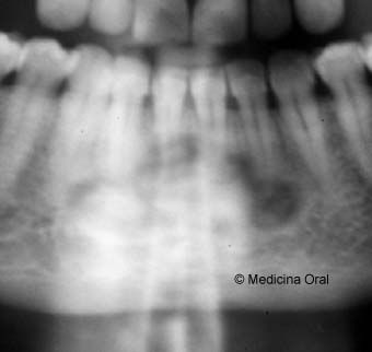
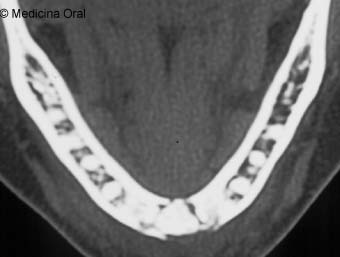
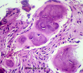
Paciente mujer de 49 años que de forma casual en una exploración radiográfica se le detectó unas alteraciones en mandíbula, zona central de incisivos. 

No tomaba medicamentos ni padecía enfermedad sistémica alguna. 

Los dientes próximos a la lesión mandibular eran vitales (pulpometría).No ha tenido alteraciones inflamatorias ni infecciosas en esa área. 

Se realizó además de la ortopantomografía en la que se visualizaron unas zonas radiolúcidas y radiopacas alrededor de los ápices de los dientes inferiores del grupo incisivo  (Figura 1), un TAC  (Figura 2)y una biopsia mandibular  (Figura 3)


SYMPTOMS AND CLINICAL EXAMINATION 


On routine examination, a 49 year old woman, was found to have a radiographic alteration in the anterior mandible in the area of the central incisors. 

The patient denied any systemic diseases and was not taking any medications. 

The teeth near the mandibular lesions were vital. The patient did not have any history of inflammation and or infectious process in the area. 

Before the biopsy orthopantomographic  (Figure 1) and CAT  (Figure 2) studies were done. (Biopsy  (Figure 3)) 


DIAGNÓSTICOS PROVISIONALES PROPUESTOS POR AQUELLOS QUE ENVIARON UN E-mail  
-Displasia cementaria periapical
-Fibroma cemento-osificante
-Displasia cemento-osificante florida
 
PROVISIONAL DIAGNOSIS PROPOSED FOR PARTICIPANTS (Those who sent an E-mail) 
-Periapical cemento-osseus dysplasia.
-Cemento-ossifying fibroma.
-Florid cemento-osseus dysplasia
 Bibliografía:
Su L, Weathers DR, Waldrom CA. Distinguishing features of focal cemento-osseus dysplasias and cemento-ossifying fibromas. I. A pathologic spectrum of 316 cases. Oral Surg Oral Med Oral Pathol Oral Radiol Endod 1997;84:301-9.


 Reference:
Su L, Weathers DR, Waldrom CA. Distinguishing features of focal cemento-osseus dysplasias and cemento-ossifying fibromas. I. A pathologic spectrum of 316 cases. Oral Surg Oral Med Oral Pathol Oral Radiol Endod 1997;84:301-9.
 
 
DIAGNÓSTICO DEFINITIVO 
DEFINITIVE DIAGNOSIS 
Displasia cementaria periapical
Periapical cemento-osseus dysplasia
 
 
© 1996-1997 Medicina Oral
Prof. José V. Bagan
UNIVERSIDAD DE VALENCIA (ESPAÑA)
VALENCIA UNIVERSITY (SPAIN)