INTRODUCTION
The type and number of
illustrations that the author can use to demonstrate ideas are
limited in traditional scientific journals. In these journals,
color printing remains prohibitively expensive and even high
quality black and white plates are expensive to produce. However
with the advent of electronic journals, currently more than 5000 (NewJour, 1997), these limitations are no longer a factor.
Authors have a host of other means to express and convey their
ideas: color illustrations, audio, animation and movies all can
be used to great effect.
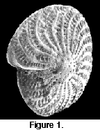 In this paper we present an exciting example
of the potential of the electronic medium in paleontological
illustration. We used Apple's QuickTime VR technology and a
scanning electron microscope (SEM), to produce a series of images
of the benthic foraminiferan Elphidium crispum Linné, 1758. These images were then combined to form a
QuickTime VR object movie (Figure 1). This type of QuickTime movie allows the
viewer to manipulate the object, and depending on the complexity
of the movie, view it from all angles (Heid 1997). The benefit of QuickTime VR is that with
proper preparation the final movie is relatively easy to
generate, and instead of just a series of static images the
viewer has the ability to examine a virtual copy of the specimen.
We are not aware of any previous combined application of
QuickTime VR and SEM in the scientific literature.
In this paper we present an exciting example
of the potential of the electronic medium in paleontological
illustration. We used Apple's QuickTime VR technology and a
scanning electron microscope (SEM), to produce a series of images
of the benthic foraminiferan Elphidium crispum Linné, 1758. These images were then combined to form a
QuickTime VR object movie (Figure 1). This type of QuickTime movie allows the
viewer to manipulate the object, and depending on the complexity
of the movie, view it from all angles (Heid 1997). The benefit of QuickTime VR is that with
proper preparation the final movie is relatively easy to
generate, and instead of just a series of static images the
viewer has the ability to examine a virtual copy of the specimen.
We are not aware of any previous combined application of
QuickTime VR and SEM in the scientific literature.
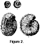 As a companion and comparison to the
QuickTime VR object movie a brief illustrative history of Elphidium
crispum is presented. Due to its common occurrence,
relatively large size (300µm) and ornate structure, Elphidium
crispum has been illustrated repeatedly in the scientific
literature during the past 300 years. The first illustrations
were drawings done in the early to middle 18th century
(Plancus 1739, Figure 2 A, B and Gualterius 1742, C, D). Generally
the poor quality of microscope optics and light sources available
made accurate representation of the species difficult. By the
late 18th century the advent of more advanced microscopy,
including the introduction of the camera lucida, permitted much
better technical illustrations to be produced (Fichtel Moll 1798,
As a companion and comparison to the
QuickTime VR object movie a brief illustrative history of Elphidium
crispum is presented. Due to its common occurrence,
relatively large size (300µm) and ornate structure, Elphidium
crispum has been illustrated repeatedly in the scientific
literature during the past 300 years. The first illustrations
were drawings done in the early to middle 18th century
(Plancus 1739, Figure 2 A, B and Gualterius 1742, C, D). Generally
the poor quality of microscope optics and light sources available
made accurate representation of the species difficult. By the
late 18th century the advent of more advanced microscopy,
including the introduction of the camera lucida, permitted much
better technical illustrations to be produced (Fichtel Moll 1798,
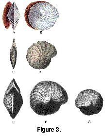 Figure
3 A, B; Williamson 1858,
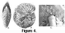
Figure 3 C, D; and Brady 1884, Figure 3 E, F in Barker 1960). The epitome of static illustration was
achieved when K. C. A. Smith of the Cambridge University
Engineering Laboratory in England constructed a SEM for the Pulp
and Paper Research Institute of Canada in 1960. This SEM was the
first that was capable of producing images of present day quality
(Wells, 1974). This equipment permitted excellent
quality images, often at high resolution and resulted in an
explosive increase in micropaleontological research (Figure
4 A-4C ). The advent of
QuickTime VR technology, coupled with SEM images, has the
potential to revolutionize micropaleontological illustration
again.
Figure
3 A, B; Williamson 1858,
Figure 3 C, D; and Brady 1884, Figure 3 E, F in Barker 1960). The epitome of static illustration was
achieved when K. C. A. Smith of the Cambridge University
Engineering Laboratory in England constructed a SEM for the Pulp
and Paper Research Institute of Canada in 1960. This SEM was the
first that was capable of producing images of present day quality
(Wells, 1974). This equipment permitted excellent
quality images, often at high resolution and resulted in an
explosive increase in micropaleontological research (Figure
4 A-4C ). The advent of
QuickTime VR technology, coupled with SEM images, has the
potential to revolutionize micropaleontological illustration
again.