![]()
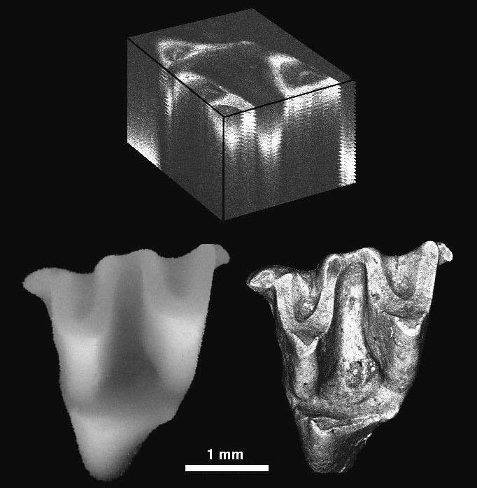
Figure 3. Digital elevation model (lower left) and maximum brightness image (lower right) generated from an image stack (top and Fig. 2). The DEM gray levels correspond to height. The image stack on the top is made of confocal images at 50 µm intervals. Note that the stack (top) is cut open to show a part of the tooth. Anterior (mesial) is toward the left, and lateral (buccal) toward the top.