![]()
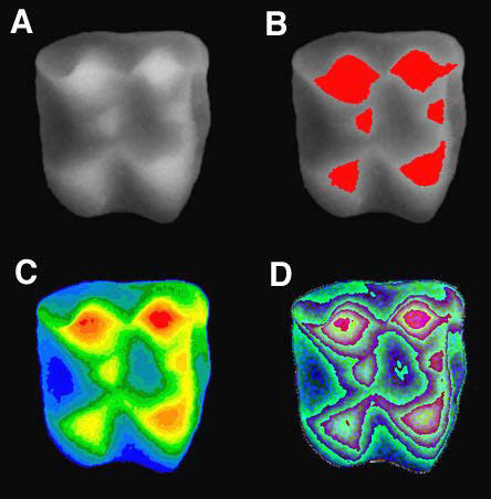
Figure 6. Digital elevation model A. of the echinosoricine molar from Figure 5 showing different ways to color-code the shape. Cusps can be easily delineated B. based on their height information revealing the relative size of cusps. In false color images, each gray-level is replaced with another color C. so that the separation of cusps and crown base becomes more apparent, or repeated color gradients (D) can aid precise recognition of landmarks (e.g., cusp tips) for measurement. Anterior (mesial) is toward the right, and lateral (buccal) toward the top.