.svg)


Dicovery of the changes that the brain suffers long before the first symptoms of Alzheimer
- Scientific Culture and Innovation Unit
- April 12nd, 2019
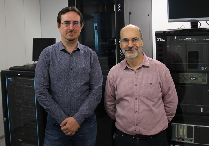
De izquierda a derecha: José Vicente Manjón (UPV) y Enrique Lanuza (UV).
A research team of the Universitat de València, the Universitat Politècnica de València, The National Centre for Scientific Research of France and the University of Bordeaux have developed a model which offers new clues about the evolution of Alzheimer. The study determines that the neuropathological changes start much earlier than the clinic symptoms and years before the clinic diagnosis. The affected by this pathology suffer an atrophy of the hippocampus between the 37 and 39 years old and an atrophy of the tonsil between the 40 and 44 years old.
There is a consensus in the scientific community about the early start of Alzheimer, well before the apparition of the first symptoms. Now, a research of the Universitat de València (UV), the Universitat Politècnica de València (UPV), the National Centre for Scientific Research of France (CNRS is its acronym in French) and the University of Bordeaux (UB) has described the changes that specific brain structures suffer along a lifetime, and allows to predict the moment in which the trajectories of the model pathological and the non-pathological separated from each other.
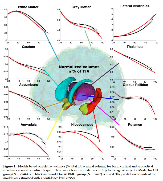
The research, developed by Enrique Lanuza (UV), José Vicente Manjón (UPV), Pierrick Coupé (UB) and Gwenaelle Catheline (UB) and published in the Scientific Reports magazine, has shown a divergence around the 40 years old in the volume of the hippocampus, the tonsil and the lateral ventricles of the Alzheimer's model, in comparison with the model of regular ageing. In the case of the hippocampus and the tonsil, the volume decreases meanwhile in the case of the lateral ventricles the volume increases in the pathological model.
The hippocampus is the brain structure which shows the more early divergence between the cognitive regular model and the pathological model, being detectable between the 37 and 39 years old depending on the cognitive impairment. For its part, the tonsil is the part which experiments more changes in proportion to its size in the moment of divergence between the 40 and the 44 years old. This deviation is not surprising, since the responsible for the impairment of the capacity of processing emotions and, probably, it is also related with the olfactory deficits, frequent symptoms in the patients with Alzheimer.
Regarding the model of lateral ventricles, the research show that exists a early divergence between the 39 and the 42 years old. However, the implication of this structure during the regular ageing reduces the anomaly after the 60 years old. Thus, the use of this biomarker is difficult for cases of late start beginning of the disease, since the enlargement of the lateral ventricles is produced during the regular ageing.
According to Enrique Lanuza, researcher and professor of the Department of Cellular Biology, Functional Biology and Physics Anthropology of the Universitat de València, ‘these results suggest that neuropathic changes underlying Alzheimer begin before the apparition of clinic symptoms and years before the clinic diagnosis.’
Reference framework to understand Alzheimer’s evolution
This work stablishes a reference framework which allows understand which is the dynamic of a healthy brain and which is the dynamic of a brain that suffers Alzheimer. To reach this statement, the research team analysed 4000 images of Magnetic Resonance of healthy and ill brains from subects aged between 9 months and 94 years old. For this, they used ‘volBrain’, a free platform developed by the team of the UPV and the CNRS which allows doing an automatic, fast and detailed analysis of the volume of different brain structures.
In the study 2944 resonances were evaluated, of healthy brains, from which they developed the model of the regular evolution of the cerebral volumes in a lifetime and with other 1400 patients with Alzheimer with more than 55 years old, it was built the model of sick brains. ‘From the comparison of both models, the study allowed to determine when these changes take place on the brain’ highlights José Vicente Manjón, researcher of the group IBIME-ITACA from the Universitat Politècnica de València
Creation of drugs
The researcher of the UPV remarks that apart from this information, this work could help in the creation of drugs to slow down the progress of the pathology. ‘The new model proposed gives us information about the changes in the brain in an early phase (from 40 to 55 years old) of the illness and from which we still did not have data. This opens the door to study the effect of future drugs in a preclinical phase when the deterioration of the brain can still be reversible’.
About volBrain
volBrain offers information about the volumes of the cranial cavity tissue (ICC) as well as about some macroscopic areas as the cerebral hemispheres, the cerebellum and the brain stem. It also provides the volumes and index of asymmetry of the subcortical structures, with great importance in the neurological field. All of this information is relevant for the progress of research on neurological pathologies.
Among its main advantages, volBrain stands out for its ease of use and analysis of speed, which makes it different from similar systems that exist on the market. Currently, the system has processed more than 130.000 brains all around the world and processes an average of 4000 cases per month. In the research have cooperated the National French Agency of Research and the Ministry of Economy, Industry and Competitiveness.
Article:
Pierrick Coupé et al. «Lifespan Changes of the Human Brain in Alzheimer’s Disease». Scientific Reports volume 9, Article number: 3998 (2019)


File in: Recerca, innovació i transferència , Internacionalització recerca , Investigació a la UV , Grups de recerca , Facultat de Ciències Biològiques , Organització , Cultura Científica , Difusió i comunicació científica , Finançament recerca
Other News


Tags
Covid
Culture
Studies
Institutional
Organisation
People
Research, innovation and transfer
University and Society
Living at the University