.svg)


Universitat de València and INCLIVA research centre develop new technology to improve cancer diagnosis
- Science Park
- March 9th, 2020
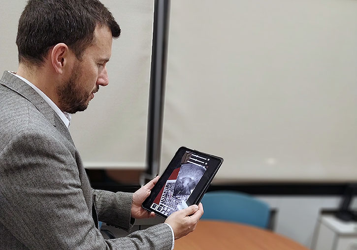
Pedro Morillo using a Mixed Reality tool designed by the UV
The UV Research Institute on Robotics and Information and Communication Technologies (IRTIC) and the INCLIVA research centre will apply an interaction paradigm system of virtual reality (VR) to improve the accuracy of interpretation of CT scans. This technology will increase the surgery success rate in cancer patients and reduce the costs and time of treatment.
Coordinated by an IRTIC researcher Pedro Morillo (Computer Science Department of the Universitat de València), the study aims to tackle one of the challenges of the RIS3CV strategy (Research and Innovation Smart Specialisation Strategy) suggested by the Valencian government (Generalitat Valenciana) and implemented by Agència Valenciana d'Innovació (AVI - the Valencian Innovation Agency).
This technological solution is based on the scientific application of the VR paradigm to visualise an CT scan and, thus, help medical practitioners interpret it.
To diagnose cancer, a patient currently has to undergo a computed tomography (CT) along with other medical tests. Only after interpreting a CT scan, doctors may decide upon the necessity of a surgical intervention.
This visualisation form complicates the spatial understanding of the CT scan; therefore, its analysis and interpretation is a tiring process that results in an error probability of more than 20%. In other words, at least 20 out of 100 patients may be mistakenly diagnosed with operable cancer if the real tumour location and anatomy is studied in the process of a surgery.
A mistake in the diagnosis calling for a surgical intervention may result in a double damage for the patient. First, an unnecessary surgical intervention presupposes anaesthesia, postoperative recovery, and so on. Second, it often means a delay in a more suitable for a patient treatment.
The project offers another solution: to apply the VR interaction paradigm to improve the accuracy of interpretation of CT scans, which will lead to higher success rates in diagnosis and lower costs and time of cancer treatment. This new visualisation method not only will be compatible with a CT screening but also with 3D images and, therefore, can complement current working conditions without losing any diagnostics options available until now.
The new system will allow the medial practitioners to visualise the information of the tests as if they were directly observing one´s internal organs and will offer all the necessary tools to interact with the abovementioned information.
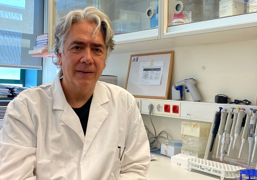
The design of this technological solution was possible thanks to the experiments and validation procedures of the team supervised by Luis Sabater, a professor at the Department of Surgery of the Universitat de València, a director of the Hepato-Pancreatico-Biliary Surgery Section of the University Hospital Clinic of Valencia, and a member of the INCLIVA Scientific Management. This team is a pioneer in the use of 3d models of tumours and livers to plan extreme oncological surgeries.
To fund the project, the researchers participate in the 2020 call “Value and Transfer of the Research Results” of the AVI - the Valencian Innovation Agency. Pedro Morillo claims that “this project is a little proof of the transfer capacity of the Valencian innovation.” Morillo pointed out that many fruits of cooperation between the Research Institutes and the industrial sector of Valencia result from the challenges posed by the AVI Innovation Strategy Committee.
File in: Ciencias Tecnológicas , Ciencias Médicas

Tags
Covid
Culture
Studies
Institutional
Organisation
People
Research, innovation and transfer
University and Society
Living at the University