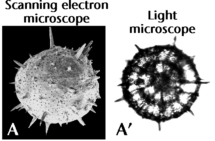
| Figure 5A, A’. Scanning electron microscope (left) and light-microscopy (right) photographs of the same radiolarian specimen, Cromyechinus antarctica. Notice that while SEM pictures yield great details of the surface of the shell-wall, they conceal all internal structures, most of which are important for identification purposes. From Boltovskoy (1981e), and Boltovskoy et al. (1983). |
 |