

with Gottron’s papules
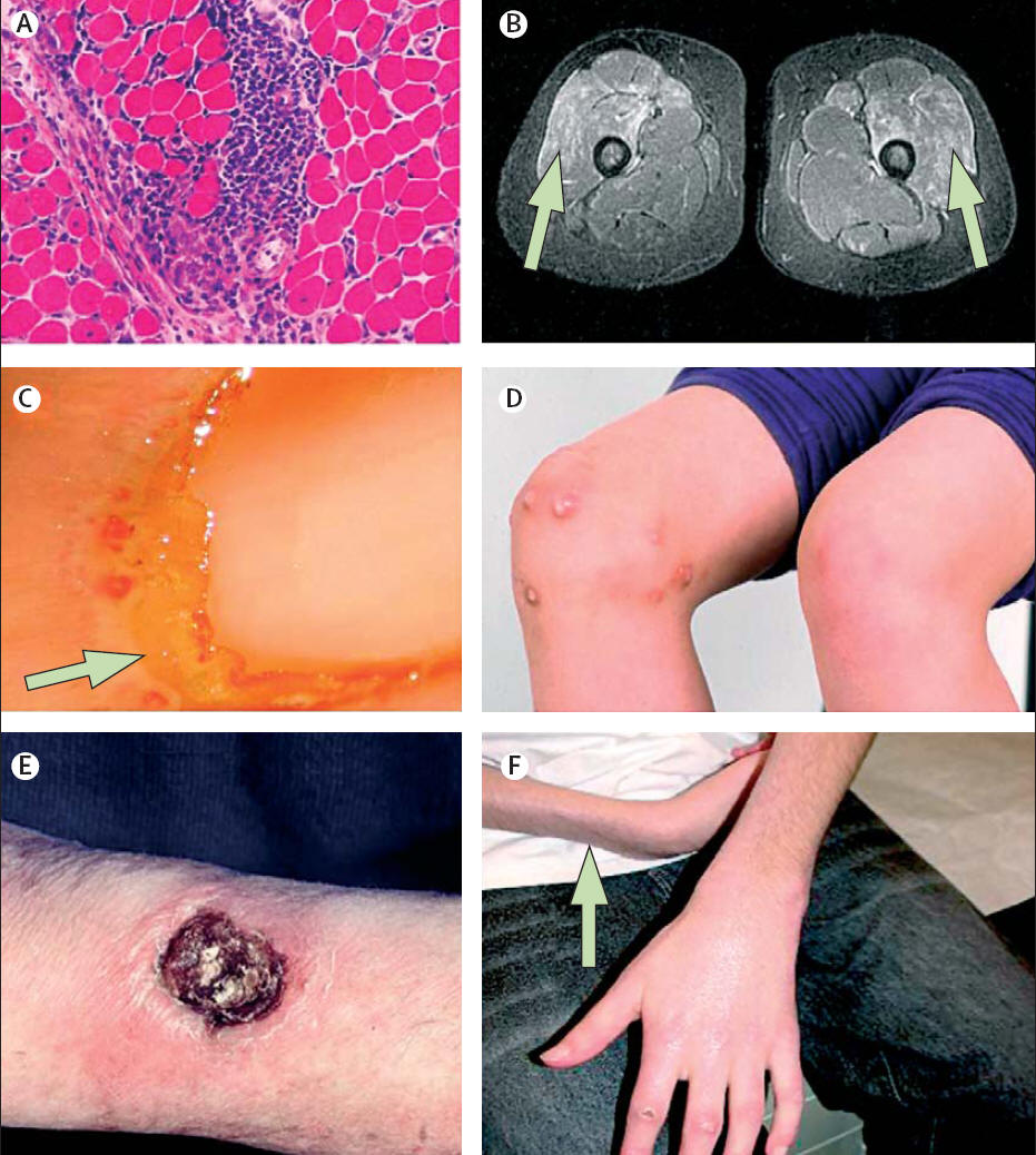
perifascicular atrophy and regeneration in a muscle biopsy. (B) MRI is a sensitive indicator of myositis. Infl amed
areas appear bright on short-tau inversion recovery-weighted images (arrows). (C) Capillaries are most often
abnormal when viewed at the nailfold. Typical changes of dilatation with adjacent drop out (arrow) is seen.
(D) About 30% of JDM patients have dystrophic calcinosis. (E) Cutaneous ulceration with central necrosis, crust and
surrounding erythema at the elbow of a 10-year-old boy with severe JDM. (F) Lipoatrophy of the forearm (arrow)
in a boy with JDM.
infl ammatory myopathies of childhood. www.thelancet.com Vol 371 June 28, 2008