Structured illumination Microscopy
|
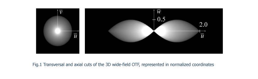
Conventional light microscopes have certain limitations regarding to the transmitted spatial frequencies both in the transverse as in the axial direction. These restrictions are imposed by the structure of the three-dimensional optical transfer function (OTF) which relates the transmission of the spatial frequencies from the object irradiance distribution to its corresponding image. The OTF has a certain bandwidth in its transversal cut showing a cut-off spatial frequency which determines the resolving power of the system. This is the physical limit imposed by diffraction for an imaging system under conventional illumination. Moreover, the lengthwise section of the OTF presents a region of missing frequencies, known as the missing cone, which implies that there is no transmission of axial spatial-frequencies through the imaging system. This missing cone is the responsible for the lack of optical sectioning capability of conventional microscopes. In Fig.1 we have represented, in normalized spatial-frequencies, the transverse and meridian cuts of the 3D OTF for a clear circular aperture.
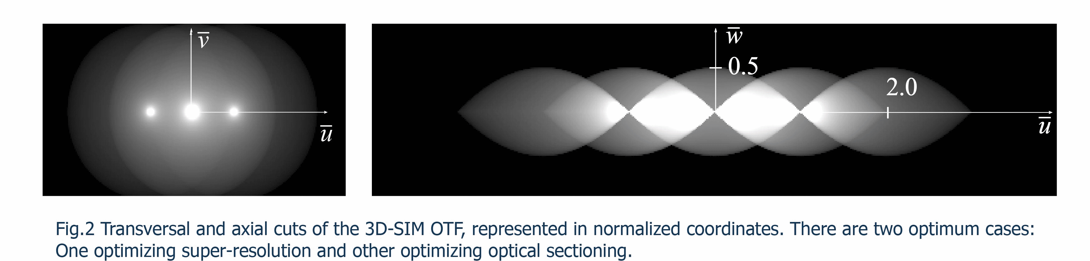
As has been demonstrated in recent years, it is possible to overcome these limitations by using non-conventional illumination methods. One of these techniques, known as structured illumination (SI), permits to surpass the classical resolution limit and provides optical sectioning. This technique is based on the projection of a one-dimensional periodic illumination pattern onto the sample. Due to the periodicity of the pattern, the illumination acts as a carrier wave which encodes different parts of the object spectrum in the spatial frequency domain. An enhanced-bandwidth OTF is reconstructed by decoding the information which is added to the object spectrum. This procedure requires the acquisition of several images (at least three) shifting properly the illumination pattern along the transversal direction. Then a synthetic image is computationally obtained by means of a reconstruction algorithm. Depending on the frequency of the projected pattern it is possible to achieve superresolution in the transversal direction or optical sectioning capability. In Fig.2 we have represented the OTF in both cases.
The conventional way to project a SI pattern onto the sample is shown in Fig.4. Bassically, a 1D grid diffracts a collimated-monochromatic beam. Then, two plane waves are generated and projected by means of a microscope objective within the specimen. The light reflected by the sample (or emmited in the case of fluorescence response) is collected by the same objecive lens. Taking into account that a lateral shift of the grid produces a phase-shift in the illumination pattern, images are taken when shifting the pattern and then combined to produce a synthetic image. For a given optical elements of the system, the spatial-frequency of the SI illumination depends on the perior of the grid. As a consequence, for different objective lenses or well for optimizing super-resolution or optical-sectioning various grids are needed.
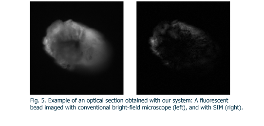
In our group, we investigate for new ways of producing structured illumination. Concretely, we have built a SI microscope in which the frequency of the projected pattern is easily switchable. Moreover, our system can be conducted with both coherent and incoherent light. This system is based on a very simple concept, which is illuminating a 1D diffracting element with a point source. Starting from that point, we have developed a motorized system with its corresponding software, being able to obtain super-solved images and z-stacks with low acquisition time. The system works also as a optical-sectioning microscope. By using other algorithm, SI technique can be applied in order to achieve slices of the sample. This algorithm, based on heterodyne detection, is used as a form of demodulating the sinusoidal illumination. If this illumination is rapidly defocused (incoherente case), then it is possible to retrieve the information of the zone in which the pattern is contained, neglecting the background light, as it can be seen in Fig.5. In this figure we show the images (60x,NA=0.8)of a fluorescent bead corresponding to wide-field and SIM acquired with our system.
|