|
PSF-Engineering
|
|
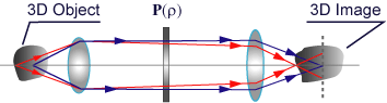
One of the challenges in three-dimensional microscopy is to overcome the lack of isotropy of the spatial resolution, which results from the axially-elongated shape of the point spread function (PSF). Such anisotropy gives rise to images in which significant axially-oriented structures of the sample are not resolved. On the other hand, the beam emerging from the high-NA objective focuses deeply through an interface between two media of different refractive index. Such a refractive-index mismatch introduces an important amount of spherical aberration, which increases dynamically when scanning at increasing depths. This effect strongly degrades the 3D PSF, and therefore the instrument performance. Quasi-isotropic 3-D resolution The use of wide-field conventional optical microscopes to image 3D biological fluorescent samples has the drawback that any image focused at a certain depth in the sample contains blurred information from the rest of the sample. This fact gives rise to 3D images with deteriorated contrast and SNR.
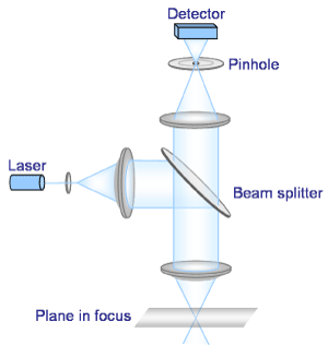
Figure 1.-Schematic geometry of a wide-field conventional optical microscope To overcome this problem, the use of scanning fluorescence techniques has been demonstrated and routinely used. Among them we cite first single-photon confocal microscopes (SPCM). The main advantage of SPCM is their sectioning power, due to the light rejection from out-of-focus parts of the sample.
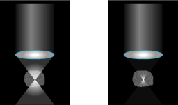
Figure 2.-Schematic geometry of a confocal microscope Serious drawbacks of SPCM are photo-bleaching, which occurs since the entire sample is bleached when any single plane is imaged, and its poor depth-penetration capacity. To solve these problems, two-photon excitation (TPE) scanning microscopy was demonstrated. This non-linear imaging technique relies on the simultaneous absorption of two photons, whereby a single fluorescence photon is emitted. The overall fluorescent light is collected and finally the image is synthesized from the 3-D sampling of the object. TPE generally uses near-infrared light due to its lower absorption and scatter in biological tissues, which allows deeper penetration of the excitation beam. As shown in Fig. 3, in TPE photo-bleaching is restricted to the neighbourhood of the imaged plane.
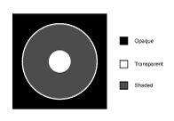
Figure 3.- In TPE fluorescence processes (right-hand figure) the bleaching is restricted to a small regions surrounding the focus We concentrate on apodization of the TPE mode with the aim of reducing anisotropic imaging, by pupil spatial filtering with a shaded-ring filter (SR), Fig. 4. We demonstrate that a close to spherical 3-D PSF can be obtained through the implementation of a relatively inexpensive spatial filter.
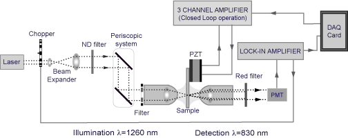
Figure 4.- The SR filter. The transmittance of the shaded ring is 19% In our experiment we used as the illumination source a mode-locked Cr4+: fosferite laser. The laser produced pulses with central wavelength l=1260 nm and a mean temporal width Dt=50 fs at a repetition rate of 84 MHz. An average power over 200mW was available for the experiment. We set the coordinate system so that the incident light is seen as x-polarized. For the illumination and collection we used two identical 1.2 NA (water-immersion) objectives from Olympus (Uplan APO/IR 60x). In Fig. 5 we show a scheme of the experimental setup.
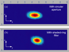
Figure 5.-Schematic geometry of the TPE scanning microscope In Fig. 6 we present typical results for the PSF measurements obtained with and without the SR filter. The PSF obtained with the SR is clearly narrowed along the optical-axis direction, thus exhibiting close to spherical shape.
Figure 6.- 2D sections of the experimental 3-D PSFs as displayed by LabView Quasi-isotropic 3-D resolution can be produced in TPE scanning microscopy by spatial modulation of the illumination beam, which involves a small modification of the microscope architecture. Reduction of spherical-aberration impact
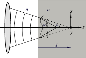
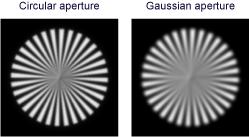
Figure 7.-Focalization of the light through one interface It is well known that, in a paraxial focusing regime, an aperture stop with Gaussian transmittance allows the production of a focused spot whose form changes very slowly with the spherical aberration (SA). However, a Gaussian pupil is not a realistic solution for the SA problem in high-NA scanning optical instruments. This is because, the Gaussian aperture is nothing more than a reduction of the effective NA of the objective lens. In Fig.8 we show how the cut-frequency is much bigger in the case of the circular pupil than for the Gaussian aperture, both in absence of SA.
Figure 8.-Numerically evaluated images of a typical spoke target, in absence of spherical aberration To take profit from the abilities of Gaussian transmittances, but avoiding the reductions in effective NA, we find another pupil, the reversed-Gaussian aperture, with identical response to SA, but opposite transverse response than the Gaussian pupil.
Figure 9. Amplitude transmittance of the reversed-Gaussian aperture We have calculated the intensity distribution in the best focal plane for both cases: the circular and the reversed-Gaussian aperture. As we see in Fig. 10, in absence of spherical aberration the circular aperture still produces slightly better results. However, as the SA increases the response of the circular aperture degrades very fast, whereas the response of the reversed-Gaussian profile remains fairly unchanged.
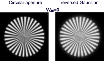
Figure 10.- Lateral responses corresponding to a high-NA focusing element with the circular (black line) or the reversed-Gaussian aperture (red line) as the aperture stop, and for increasing values of w40 To have a visual insight of the improvements obtained with the reversed-Gaussian aperture we have performed a numerical imaging experiment. The images were obtained by 2D convolving the lateral responses in Fig. 10 with the spoke target. The results are shown in Fig. 11.
Figure 11.-Numerically evaluated images of a typical spoke target, for increasing spherical aberration. Note that in absence of aberrations the performance of the circular aperture is slightly better. However, the reversed-Gaussian aperture is much more robust against the increasing of the SA. In other words, we can affirm that the reversed-Gaussian profile confers the high-NA system an important tolerance to the SA. This tolerance allows the high-NA optical instrument to have a performance that is not degraded as scans deeply into a medium of different refractive index.
|