

The challenging lecture given in 1959 by physicist and Nobel Prize awarded R. P. Feynman: “There's plenty of room at the bottom” is considered to be the starting point for nanotechnology
With this peculiar title, Feynman encouraged researchers to explore beyond the atomic level and predicted exciting new phenomena that might revolutionize science and technology. Among these pioneering researchers are Eric Betzig, Stefan W. Hell and William E. Moerner, who have been awarded with the Nobel Prize in Chemistry 2014 for developing the super-resolved fluorescence microscopy. However, it is important to remark that the exploration of this amazing nanomolecular world began in the early 1980s with the invention of the scanning tunneling and the atomic force microscopes.
The study and manipulation of interactions of nanometric dimensions could begin as soon as measuring tools became more efficient. The last decades have witnessed the development of techniques able to obtain information at the sub-molecular level, which have been applied mainly to the biomedical field. As an example, the stimulating labor of studying the role of conformational dynamics in reaction mechanisms has resulted in numerous advances in life sciences. The precise knowledge about molecular interactions and their effects on protein function has greatly aided the discovery of new targets in medical chemistry. Thus, the ability to provide information linking these different areas is of fundamental interest. Usual techniques span from those providing exquisite details in terms of molecular structure of a static system, such as X-ray crystallography or neutron reflection (NR), to those providing dynamic measurements, which include electrochemical and optical techniques. However, the measurement of conformational dynamics of chemical processes displayed in a label-free and real-time manner is no longer reached by few ways.
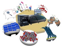
Dual Polarization Interferometry (DPI) is currently one of the most powerful techniques for recording real-time data of conformational dynamics. It is a novel tool with current applications in surface science and biophysics. This technique is, to our knowledge, the most well-thought through such waveguide sensor. It has its weaknesses, e.g. the necessity of using a relatively long sensor element. However, it is well known that the use of an interferometric readout and a long waveguide makes the measurements very stable and more accurate than other techniques, such as quartz crystal microbalance or surface plasmon resonance. Accordingly, DPI can be described as a molecular ruler whose quantitative values can be correlated directly with those from other usual techniques, such as NMR, X-ray crystallography and NR, providing higher sensitivity and accuracy than the classical acoustic and optical biosensors. Swann et al. (Swann, M. J.; Peel, L. L.; Carrington, S.; Freeman, N. J. Anal. Biochem. 2004, 329, 190) proposed an elegant assistant for the interpretation of DPI data by correlating the different parameters (thickness, density, mass coverage) calculated, in real-time, from the interferometric detection of optical field phase changes.
The key strengths of DPI are, in first place, the use of untagged reagents, which clearly simplifies the number of steps of the experimental procedure. Secondly, the short response time, which allows data updates at least every 20 ms, provides quantitative data on real-time changes in dimension, refractive index and density and thirdly the high sensitivity with resolution below 0.1 Å, 10-7 refractive index units and 0.1 pg/mm2, respectively. For all these reasons, this technique offers a unique perspective on biochemistry, linking conformational changes to biologic activity at a resolution usually attributed to ‘big physics’. Additionally, DPI has three important features that offer a different approach in the investigation of biomolecules structure change, such as:
- Full structural characterization of every system in real-time (thickness, refractive index, surface concentration, density, area per molecule and volume fraction). This high level of quantitative characterization allows monitoring real-time actions of biomolecules which can be resolved as regard shifts in the layer’s dynamic structure.
- Accurate measure of birefringence, which allows the changes in the supported anisotropic layer’s structure to be measured in real-time, which results in a better knowledge of the organization effects. These studies have important applications in lipidomics.
- Kinetic analysis of the binding process. This means that structural alterations due to binding can be monitored in real-time, providing an incredible insight into the binding mechanism thanks to the technique performances.
It is important to emphasize that DPI is emerging as a viable, rapid and highly sensitive technique that holds great potential in becoming a standard tool in functional biological studies and characterization for a wide variety of applications. It is important to note the significant increase of the DPI use in the last years. It is possible to predict that this technique can become a basic tool for studying biomolecular interactions and surface characterization. It can be an essential reference for the expected growing of research efforts in conformational dynamics. Accordingly, most examples included and discussed in a recent review correspond to multidisciplinary publications appeared in the period of existence of the technique (http://pubs.acs.org/doi/abs/10.1021/cr5002063 ). This review discusses the basic aspects of the exciting, tagless DPI technique and provides an update on its current and potential applications. All this gives a unique chance to learn from a novel tool.


