CIGE/2021/167 Automated Cromatic Contrast Sensitivity Function
Development of a new psychophysical test to measure contrast sensitivity to chromatic and achromatic stimuli to evaluate functional losses in visual pathologies.
Acronym
FSCA
Description
SCIENTIFIC PROPOSAL
1. Background
The contrast sensitivity function (CSF) is a psychophysical measure that allows the detection of a wide variety of ocular and systemic pathologies even in early stages of pathology evolution.1-4
The most common devices in clinical practice focus exclusively on the evaluation of the achromatic CSF (see section 1.2). However, the analysis of the sensitivity of the opponent chromatic mechanisms (red-green and blue-yellow) is relevant, because, although in many cases it is not possible to ensure that a certain pathology affects preferently a single visual mechanism, it has been shown that functional losses linked to anatomical losses are detected earlier in some mechanisms than in others.2 ,4-6
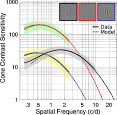
The achromatic CSF acts as a band-pass frequency filter and the CSFs of the chromatic mechanisms are low-pass, with the red-green mechanism being more sensitive, especially at low frequencies (see figure 1).7 The high sensitivity of the achromatic CSF in the range of high spatial frequencies makes it difficult to measure chromatic contrast sensitivity in clinical settings, since it must be ensured that the stimuli used contain a pure color change, without luminance changes (isoluminant stimuli). A small residual achromatic signal in the chromatic stimuli can be easily detectable by the achromatic mechanism, and the resulting curve would therefore not reflect the state of a chromatic mechanism. Determining an individual equiluminance condition for each patient (and, theoretically at least, for each frequency) can be difficult for a patient without prior training and lengthens the measurement, both undesirable conditions in a clinical setting.
Figure 1. Contrast sensitivity functions for the achromatic (black line) red-green (red) and blue-yellow (blue) mechanisms7.
2. State-of-the-art and justification of the proposal
The variations of the achromatic CSF with age are well documented8, but the same does not occur with chromatic CSFs, where we find studies that include patients of various ages, but we have only found one study that evaluates, with few patients, the influence of age on chromatic contrast sensitivity for the red-green mechanism.9 However, it has been found that the sensitivity of both chromatic mechanisms decreases with age.10-12
Furthermore, we have not found studies where the gender perspective is included in their methodology, so the influence of gender on the perception of chromatic contrast has not been studied.
There are several devices to measure sensitivity to achromatic contrast, in clinical practice,, the FACT (Functional Acuity Contrast Test) being the most used,13 but we did not find any that allow us to measure the function of sensitivity to chromatic contrast. To assess the response of the visual system to red-green and blue-yellow chromatic contrasts, some members of the research team designed an experimental device (ATD Multichannel Perimeter) capable of favoring the response of each of the two visual chromatic mechanisms and that has already been used for this purpose as a perimeter.14-16 But the device used is bulky (CRT monitor) and its transportation and operation are not very ergonomic. Furthermore, it has the drawback that the perimetric measurements are very long and the chromatic stimuli are not isoluminant for each individual patient, which can lead to the intrusion of the achromatic mechanism in the response.
To perform clinical measurements of CFSc, it would be desirable to have a device 1) based on stimuli and tasks that minimize the possibility of intrusions of the achromatic mechanism without requiring the precise determination of the isoluminance condition, 2) with reduced dimensions, which ensure its portability and reduce its cost, like a tablet, 3) with a psychophysical method that reduces measurement time17 and that includes an easy task that can be performed by people with functional diversity, without compromising reliability and 4) that has a normal database for different age ranges that includes the possible effect of gender.
3. Aim
3.1. Main aim
To develop a low-cost digital device to measure the sensitivity function to chromatic contrast of both the red-green and blue-yellow mechanisms, usable for diagnosing visual disorders in a wide age range.
3.2.Specific aims
- To colorimetrically characterize two low and medium end tablets that will be used to generate the stimuli for the measurement.
- To study the optimal configuration of the tablets and the measurement conditions, with particular attention to the optimal measurement distance, considering the spatial resolution of the tablets and the range of frequencies that would be desirable to generate.
-To implement an application for a mobile device that allows rapid and reliable measurement of cCSF and perform analysis of the result and diagnosis of the patient.
-To carry out a repeatability study of the measurements with real observers.
-To obtain the reference standard observer for diagnosis, from the measurements of contrast sensitivity functions with healthy patients aged between 20-70 years.
4. Methodology and workplan
The project consists of 5 phases:
1st phase: device setup
2nd phase: generation of stimuli
3rd phase: device validation
4th phase: measurement of CSFs for different age ranges
5th phase: analysis of results, dissemination and publication
The first phase will consist of optimizing the configuration of the device's components. The tablets that will be used must be correctly calibrated and characterized for good control of the stimuli that will be generated. To do this, different stimuli generated on the tablet will be measured with a spectroradiometer. With these data, the ICC profile of the device will be calculated (response relationship between the digital levels and the luminance of each type of RGB pixel), which will allow the generation of the stimuli to be generated for the tests to be colorimetrically controlled. Reproduction errors will be estimated with the derived characterization models, and if they are not acceptable, characterization by interpolation tables (look-up table, LUT) will be used. A spatial calibration of the stimulus will also be carried out and the range of observation distances to correctly display the stimuli and all the necessary spatial frequencies will be determined.
The difficulty of this phase would be that with a low-cost tablet we do not achieve a good reproduction of the generated stimuli.
The contingency plan is to buy two tablets, one low-cost and the other medium-cost with better features to be able to compare the results.
In a second phase, a chromatic contrast sensitivity measurement test will be implemented in the MATLAB® environment.
First, a test will be implemented to evaluate the equiluminance condition of each observer, so that we avoid the intrusion of the achromatic mechanism. The test will consist of two alternating stimuli with a high flicker frequency. The observer can vary the luminance ratio of the test so that when the flicker disappears, both stimuli will have the same luminosity for the observer (Heterochromatic Flicker Photometry). The stimuli will be generated in the cardinal directions of the opponent modulation color space.14 Sinusoidal gratings subtending 5º for the observer with spatial frequencies of 0.5, 1, 2, 4, 8 and 16 cpg shall be used.
The background used will be an achromatic color with a minimum luminance of 50cd/m2. This value must be optimized to desensitize the achromatic mechanism, without reducing the range of colors that can be generated in the directions of the chromatic mechanisms.
The psychophysical method to measure the contrast threshold will be an adaptative method. The traditional staircase method will be compared with the fast methods used in the measurement of achromatic CSF.17 From this threshold, we calculate the sensitivity to chromatic contrast in decibels, with Eq. 1.
(1)
where ∆Rmax,i is the maximum amplitude along the j-cardinal direction and ∆Rthres,j the patient's threshold amplitude along the same direction.
Difficulties in this phase: The duration of the isoluminance condition determination phase may not be clinically acceptable. The color resolution of tablets is typically 8 bits per channel, and it may not be possible to generate stimuli with sufficiently small contrasts to assess the contrast sensitivity of a healthy patient. On the contrary, the difficulty with pathological patients or elderly patients lies in the fact that the maximum generateable contrast may not be detectable, especially if the use of a high background luminance is favored, to desensitize the achromatic mechanism.
Contingency plan: Replace the determination of the isoluminance condition with the use of achromatic noise. The problem of the sensitivity of normal subjects being higher than the color resolution of the device can be partially solved by raising the difficulty of the task.
In a third phase, the designed device will be validated through measurements in patients.
The learning effect and short-term repeatability will be evaluated. To do this, the measurement of one of the CSFc (either the red-green or the blue-yellow one) will be carried out twice with a break between measurements of 5 minutes. The evaluation of repeatability will be carried out with at least 20 healthy patients, 10 per mechanism.
Difficulties: low repeatability between measurements
Contingency plan: change the psychophysical method to another method that improves the reliability of the test
The fourth phase consists of obtaining the normality pattern by age, every 5 years, with a minimum of 5 eyes of each sex per group.
For this phase, patients who meet the following inclusion criteria will be needed:
Healthy people of both sexes, aged between 20 and 70 years.
Reasonable collaboration that allows the performance of an ophthalmological examination.
Patients who have given written informed consent to carry out this study.
Spherical ametropias between 3 and -3 D
Astigmatisms <1D
Decimal visual acuity >0.8 with correction.
Normal trichromatic color vision, determined with the FM100h test.
Absence of ocular or systemic pathologies with ocular repercussions that could interfere with the interpretation of the study results. Thus, patients with retinopathies, glaucoma, amblyopia, cataracts, etc. are excluded.
Without the use of drugs that may currently or in the past produce visual alterations, that may produce vision loss or alterations in perception such as drowsiness.
No history of drug addiction or alcoholism.
Subjects will be informed of the tests to be performed and their duration. After explaining and signing the informed consent, the following protocol shall be applied:
Clinic history. A clinical history will be established, to check that inclusion criteria are complied with.
Measurement of best corrected visual acuity.
Selection of the eye to be examined randomly, if both eyes can be included, otherwise the eye that meets the inclusion criteria will be chosen.
Evaluation of color vision using the Farnsworth-Munsell Test administered in a lighting booth under illuminant D-65.
CSFc measurement:
With the new device, the contrast sensitivity of the two opposing chromatic channels will be measured, randomizing the order in which they are evaluated.
Measurements are carried out in a dark room, with no light source other than the tablet. The subject will view the test monocularly with its correction and a chin rest will be used to stabilize the position of the head. Achromatizing lenses will be used to reduce chromatic aberration.
The task to be performed is proposed to be pattern detection. On the screen, the patient will be shown narrow-banck stimuli of different contrast and spatial frequency, and the patient must respond using a button if they can see the stimulus or not.
In the fifth phase, the results will be analyzed, evaluating whether there are significant differences and agreement between the two measurements performed on the patients. Furthermore, it will be checked if the measured CSFcs are consistent with those published in the literature and finally it will be checked if there are correlations between the sensitivity at different frequencies of the two CSFcs, the age and sex of the participants. The results will be disseminated at conferences related to the topic and the results will be published, trying to have them published in journals indexed in the JCR.
5. References
1. Bennett CR, Bex PJ, Bauer CM, Merabet LB. The Assessment of Visual Function and Functional Vision. Semin Pediatr Neurol. 2019 Oct;31:30-40. doi: 10.1016/j.spen.2019.05.006.
2. Chen XD, Gardner TW. A critical review: Psychophysical assessments of diabetic retinopathy. Surv Ophthalmol. 2020 Aug 29:S0039-6257(20)30126-0. doi: 10.1016/j.survophthal.2020.08.003.
3. Elfadaly D, Abdelrazik ST, Thomas PBM, Dekker TM, Dahlmann-Noor A, Jones PR. Can Psychophysics Be Fun? Exploring the Feasibility of a Gamified Contrast Sensitivity Function Measure in Amblyopic Children Aged 4-9 Years. Front Med (Lausanne). 2020 Aug 26;7:469. doi: 10.3389/fmed.2020.00469.
4. Sartucci F., Murri L., Orsini C., Porciatti V. Equiluminant red-green and blue-yellow VEPs in multiple sclerosis. J Clin Neurophysiol. 2001;18:582–591
5. García-Domene MC, Díez-Ajenjo MA, de Fez MD, Luque MJ Visual fields in a chloroquine treatment 2014 Clinical optometry. 2014;6 :59—70
6. Ahmed OM, Waisbourd M, Spaeth GL, Katz LJ. Improvement in structure and visual function in patients with glaucoma: the possible key to better treatment? Surv Ophthalmol. 2020 Dec 11:S0039-6257(20)30176-4. doi: 10.1016/j.survophthal.2020.12.004.
7. Kim YJ, Reynaud A, Hess RF, Mullen KT. A Normative Data Set for the Clinical Assessment of Achromatic and Chromatic Contrast Sensitivity Using a qCSF Approach. Invest Ophthalmol Vis Sci. 2017 Jul 1;58(9):3628-3636. doi: 10.1167/iovs.17-21645.
8. Lord SR1, Clark RD, Webster IW Visual acuity and contrast sensitivity in relation to falls in an elderly population. Age Ageing. 1991 May;20(3):175-81
9. Dees EW, Gilson SJ, Neitz M, Baraas RC. The influence of L-opsin gene polymorphisms and neural ageing on spatio-chromatic contrast sensitivity in 20-71 year olds. Vision Res. 2015 Nov;116(Pt A):13-24. doi: 10.1016/j.visres.2015.08.015.
10. Kinnear PR1, Sahraie A New Farnsworth-Munsell 100 hue test norms of normal observers for each year of age 5-22 and for age decades 30-70Br J Ophthalmol. 2002 Dec;86(12):1408-11.
11. Knoblauch, K., Vital-Durand, F. and Barbur, J. L. (2001) Variation of chromatic sensitivity across the life span. Vision Res. 41, 23–36.
12. Werner, J. S. (1996) Visual problems of the retina during ageing: compensation mechanisms and colour constancy across the life span. Prog. Retin. Eye Res. 15, 621–645A.
13. Ginsburg AP. Next generation contrast sensitivity testing. In: Rosenthal B, Cole R, eds.Functional Assessment of Low Vision. St Louis: Mosby Year Book Inc, 1996:77–88
14. María Amparo Díez-Ajenjo, Pascual Capilla. Spatio-temporal Contrast Sensitivity in the Cardinal Directions of the Colour Space. A Review. J Optom. 2010; 3(1): 2–19
15. Morilla A., Antón A., Jiménez B., Rodríguez C., Martínez V., Fallon M., Capilla P., Luque M.J., Felipe A., Artigas J.M. ATD perimetry in glaucoma and ocular hypertensive patiens. A preliminar study.EVER 2007, pp-64, PS3-448.
16. Díez-Ajenjo MA, Capilla P, Luque MJ. Red-green vs blue-yellow spatiotemporal contrast sensitivity across the visual field. Journal of Modern Optics. 2011;58:1736–1748.
17. Dorr M, Lesmes LA, Lu ZL, Bex PJ. Rapid and reliable assessment of the contrast sensitivity function on an iPad. Invest Ophthalmol Vis Sci. 2013 Nov 5;54(12):7266-73. doi: 10.1167/iovs.13-11743.
Tras estudiar los errores de reproducción de diferentes monitores LED, los mejores resultados con un modelo matricial de caracterización cromática se han obtenido con una pantalla retina. Después de realizar distintas pruebas sobre el tipo de estímulos de banda estrecha (texturas y ruido aleatorio), y de probar procedimientos para silenciar el mecanismo acromático en las medidas en las direcciones rojo-verde y azul-amarillo, se ha llegado a un diseño de test de medida de sensibilidad al contraste, que, con el rango dinámico del monitor y tarjeta gráfica utilizadas, permite medir la sensibilidad al contraste de pacientes jóvenes. En una siguiente etapa, será necesario determinar si los estímulos y la tarea son adecuados para medidas en otros rangos de edad y en pacientes patológicos.