

Type: Equipment
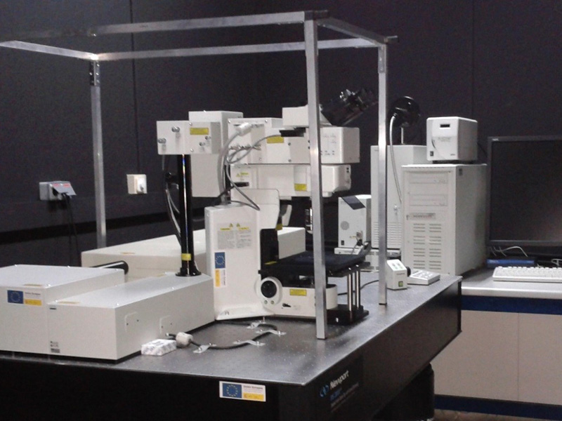
Multiphoton confocal microscope mounted on a motorised BX61WI upright microscope equipped with a XPLN25xWMP water immersion lens exclusive to multiphoton microscopy, with a numerical aperture of 1.05 and a working distance of 2mm. It has a Mai-Tai HP DeepSee pulsed tunable laser (Spectra Physics) in the 690 to 1040nm range with an average power of 2.0 W. The infrared element of this laser allows for further penetration in the sample and causes less cell damage. The signal is picked up by four detectors covering the whole visible range (420-500/515-580/590-650/660-740).
Multiphoton confocal system mounted on a BX61WI upright microscope with motorised focus and equipped with a XPLN25xWMP full water immersion lens exclusively developed for multiphoton microscopy. This lens is characterised by its numerical aperture (NA) of 1.05 with a great working distance of 2mm. This allows to go deeper into the sample with good resolution and without the risk of crushing the sample with the lens. The excitation source is a Mai-Tai HP Deep See (Spectra Physics) pulsed tunable laser in range of 690 to 1040nm with an average power of 2.0W. The fluorescence signal is detected via four photomultiplier tubes covering the whole visible range.
| PMT 1 | 420 - 500nm |
| PMT 2 | 515 – 580nm |
| PMT 3 | 590 – 650nm |
| PMT 4 | 660 – 740nm |
The multiphoton systems allow to excite the conventional fluorescent probes with a laser in the infrared range via a simultaneous photon absorption. Initially, this two-photon absorption presents certain advantages on conventional confocal microscopy:
- Higher tissue penetration, as there’s less absorption in the infrared range.
- The two-photon excitation is only produced in the focal plane, resulting in the photodegradation of the sample or fluorescent probe to be limited to a small volume.
- It enables the excitation of ultraviolet-excited probes with an infrared laser, which reduces phototoxicity when working with live cells.
- Studies for Colocalisation of Fluorescent Probes.
- Three-dimensional characterisation of biological structures and materials.
- Live cell studies.
- Microablation experiments.
ISO 9001:2015


