
.jpg)
Scientists from UV and University of Granada have developed a new molecular method that allows to define the presence of Toxoplasma gondii parasite, which causes the disease toxoplasmosis, on ham samples. Published in two articles of the scientific journal “Food Microbiolgy”, the procedure shows the protozoon’s prevalence level at 8.84% of the samples, showing significant differences between the various brands.
Toxoplasmosis is a parasitic disease that people catch basically from eating not properly cooked or handled meat, like cured meat, as well as from consuming water and vegetarian products contaminated by excrements from cats and other felines. The resulting pathology in pregnant women or immunocompromised people is important. It is estimated that 30% of world population is infected with T. gondii parasite.
The research has been carried out on 475 ham trade samples, both sliced and diced. This new method consists in capturing the parasite’s DNA by means of magnetic particles functionalised with specific sequences of the parasite’s DNA and DNA quantification by using a quantitative PCR technique (qPCR).
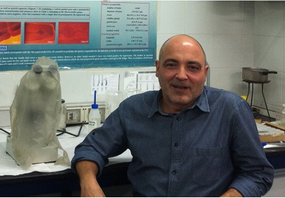
Màrius Fuentes, Professor at the UV Department of Pharmacy, Pharmaceutical Technology and Parasitlogy, highlighted the importance of “raising awareness about the intake of food which certifies the absence of the parasite, for instance, through a proper processing and curing of ham”.
The researcher underlined the security of the methodology used to define the efficacy of the curing process. He also remarked that the method would provide greater assurance for the so handled ham. Fuentes stated that “ham consumption is being questioned in some countries such as the United States. However, if the absence of Toxoplasma is certified, its purchase could increase”.
The sensitivity of this method is able to detect a parasite cell in a 100-gramme-sample of ham with 94.6% efficiency. Similarly, the scientific team specified the inefficiency of the detected parasites in the ham samples.
The team analysed 475 commercial samples of ham, presented in two formats: sliced or diced. The team estimated the number of T. gondii parasites present in the samples. This is the first study carried out on this subject.
The results showed that the prevalence level of the T. gondii in the ham samples varied from 0% until 32.35% of the samples depending on the manufacturing company. The results also indicated that 4.84% of the positive samples were ineffective on mice, that is, they were infected.
Besides, the prevalence of T. gondii in ham trade samples was of 8.84%, with a viability rate of 4.84%. Some of the samples showed high levels of both positivity and infectivity, although some of them were negative either because the parasites were not present or because they were not infectious. Analysing prevalence and viability percentage depending on the product’s type of packing (either vacuum-packed pieces or slices), no significant difference between the percentages appeared.
The authors of this research are Màrius Fuentes and, on the part of the University of Granada, Antonio Osuna, Mercedes Gómez-Samblás and Susana Vílchez, who are part of the research team CTS-183: Bioquímica y Parasitilogía Molecular (“Biochemistry and Molecular Parasitology”) of the Biotechnology Institute of Granada.
The statistical analysis of prevalence results obtained by the researchers revealed significant differences between the samples provided by different brands. Thus, parasite’s prevalence and viability in ham samples depends on the origin of the animals and the manufacturing company, which implies that the company has altered or fail to comply with the curing process’ regulations.
In the light of the results, the researchers pointed out that the use of traditional salting process for ham production ensures a total elimination of T. gondii, as long as the maturing period set by the current regulations is respected.
A danger to pregnancy
According to Professor Antonio Osuna of the University of Granada (the main author of this work), despite the health controls, the pork is still a potentially important source of T.gondii parasite, one of the worst opportunist that take advantage of immunocompromised people. It is responsible for impairments and malformations in newborns, among which we find microcephaly, hydrocephaly, blindness and congenital cardiac pathologies, if the primary infection occurs in pregnant mothers.
The researchers have proved that, when ham samples are frozen before the curing process, elimination of the parasite is much faster. Besides, they have showed that the usual ham curing process with nitrites and sea salt increases Toxoplasma survival time and infection capacity, if compared with curing process done exclusively with sea salt.
In addition, the use of nitrites during the salting process for the control of microorganisms emerged from the ham delays the T.gondii inactivation process, probably as a consequence of a delay in lipolysis phenomenon, which makes longer maturing periods necessary (at least 7 months), to ensure the total elimination of the parasite.
According to Professor Osuna, “ham manufacturers can ensure the total elimination of T.gondii by applying a freezing process on meat, either before the salting process or after the curing process.” Meat freezing processes are not established in ham manufacturing regulations, since these processes alter the organoleptic properties of some processed products, the bouque, texture and aroma of which are of high added value.
The implementation of the study could represent an advantage for the companies belonging to the manufacturing sector of ham and shoulders with an added value, given that it can ensure T.gondii free food and, therefore, their products’ security.
Captions:
1. Màrius Fuentes, Professor responsible for the UV Department of Pharmacy, Pharmaceutical Technology and Parasitlogy.
2. Image of a Toxoplasma parasite in electronic microscopy. A and B are isolated infectious forms of tissue with scanning electron microscopy. The C represents infectious tissue forms observed by transmission electron microscopy and the D shows the development of a cell’s inner part in vitro culture (IMAGE: Luis M. de Pablos Torró. The Biotechnology Insitut of UGR, Group CTS 183).
Images:


