The work, published today in the journal Nature Communications, which has used a model of rodents, has managed to relate for the first time the influence of feronomic signals on memory, which will allow in the future to explore the deficits of integration of spatial memories and social studies in transgenic models of Alzheimer’s disease. The research, directed by professors Enrique Lanuza (Faculty of Biological Sciences) and Vicent Teruel Martí (Faculty of Medicine and Dentistry), has been carried out entirely by research staff from the University of Valencia.
The evidence shows that the generation of the memory of an event or an experience includes various types of information, such as where it happened –spatial component– or who was involved –social component–, but how these components are integrated in the brain is still a mystery.
Precisely this work has managed to demonstrate that social and spatial memory are integrated into a circuit formed by three neural structures: the amygdala (part of the brain responsible for emotional reactions), the entorhinal cortex (memory and orientation) and the hippocampus (also related to memory and learning). In the case of rodents, individual recognition depends on pheromonal signals and this is the first work that shows to what extent this type of information influences memory.
According to Vicent Teruel and Enrique Lanuza, “we humans probably use the same neural circuit to integrate social and spatial information, although in our case the recognition of individuals is based on visual information instead of through the detection of pheromones by the system olfactory, as in the case of rodents”.
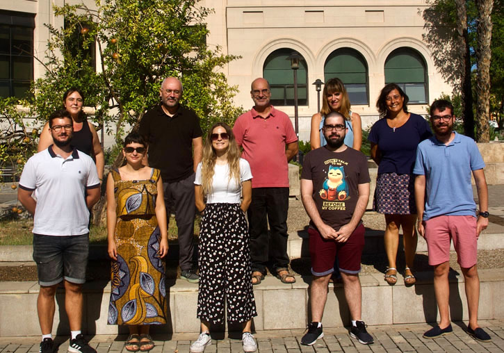
The research, funded by the Ministry of Science and Innovation-FEDER, has been carried out by María Villafranca, Esteban Merino, Ana Cervera, Joana Martínez and Vicent Teruel from the Laboratory of Neural Circuits of the Department of Human Anatomy and Embryology, and by Manuel Esteban Vila, Anna Teruel and Enrique Lanuza from the Functional Neuroanatomy Laboratory of the Department of Cell Biology. In addition, Daniel Esteve and Ana Lloret, belonging to the Department of Physiology and the INCLIVA Health Research Institute, have contributed to the work.
The methodological approach is multidisciplinary and neuroanatomical, neurophysiological, molecular biology and behaviour analysis techniques have been used, the latter using a novel open source system based on deep learning. Part of the electrophysiological recordings have been carried out in a virtual reality environment adapted for rodents, which has allowed them to virtually walk through corridors while they were presented with visual or olfactory stimuli in a very controlled way. The enormous amount of data derived from these experiments has allowed us to recognise the neural substrate of the integration of the different components of episodic memory, which will allow us to know details of this complex cognitive process.
Article:
Villafranca-Faus, M., Vila-Martín, M. E., Esteve, D., Merino, E., Teruel-Sanchis, A., Cervera-Ferri, A., Martínez-Ricós, J., Lloret, A., Lanuza, E. & Teruel-Martí, V. (2021). «Integrating pheromonal and spatial information in the amygdalo-hippocampal network». Nature Communications. https://doi.org/10.1038/s41467-021-25442-5
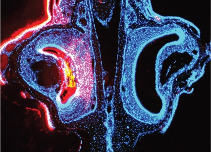
Photo caption:
Photo experiment: vomeronasal organ (responsible for detecting pheromonal signals) of the mouse. Blue: cell marker. Pink: marker of cell membranes indicating the organ activated by the detection of pheromones.
Images: