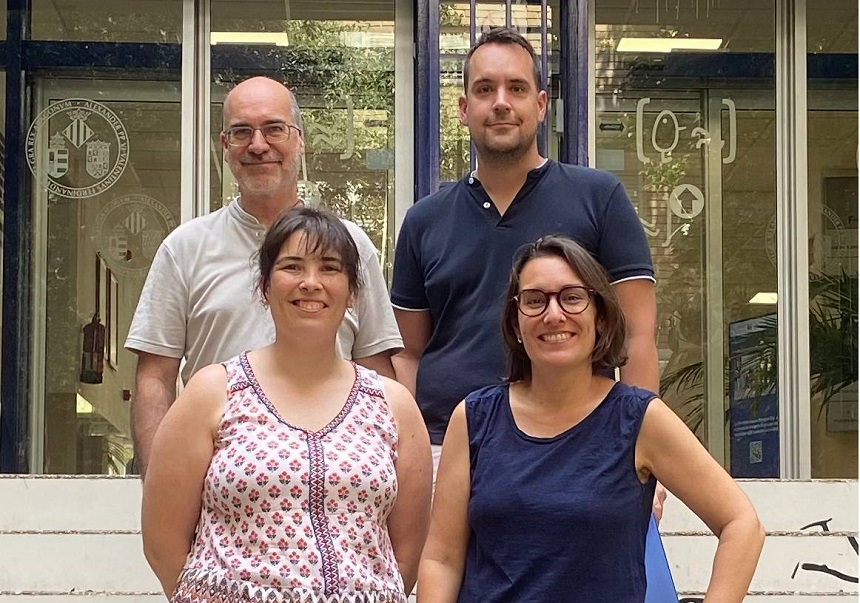
A research team from the University of Valencia (UV) has identified how early-life stress alters the morphology of immune cells in the brain, known as microglia, in mice carrying a mutation in the Mecp2 gene, associated with Rett syndrome — a rare and severe neurodevelopmental disorder that affects mainly women and causes multiple symptoms, including loss of speech and intellectual disability. The cellular changes occur in a key brain region involved in the response to pain and stress, the periaqueductal grey matter, and occur even before the first symptoms of the disease emerge.
The study, published in the journal NeuroMolecular Medicine, indicates that these morphological alterations are specific to certain brain subregions and that the microglia of MeCP2-deficient mice respond differently to stress than those of healthy mice. Microglia act as the brain and spinal cord's primary line of immune defence. These cells not only remove pathogens or cellular debris but also play a crucial role in modulating neuronal connections and the stress response. When microglia do not function properly, essential processes for brain development and functioning may be disrupted.
“The key finding is that the combination of the genetic mutation and stress at very early ages prevents microglia from adapting properly, which may contribute to the development of the typical symptoms of Rett syndrome”, explains Jose V. Torres Pérez, Ramón y Cajal researcher in the Department of Cell Biology, Functional Biology and Physical Anthropology at the Faculty of Biological Sciences of the UV and lead investigator of the study.
The research focused on female mice carrying the mutation, in pre-symptomatic stages, to study the effect of the interaction between environmental factors and genetic vulnerability. Through morphological and fractal analyses, the team observed that certain areas of the periaqueductal grey matter show altered microglia branching, while others remain unchanged.
“We observed a lack of morphological adaptation of microglia in mutant female mice exposed to early-life stress. This may reflect a failure in the brain's ability to respond adequately to adversity — a mechanism that could contribute to the alterations in pain perception or anxiety typical of the condition”, adds Maria Abellán Álvaro, first author of the article.
The study was carried out with the support of Enrique Lanuza and Carmen Agustín Pavón, also researchers in the Department of Cell Biology, Functional Biology and Physical Anthropology (UV). The work also involved authors affiliated with the Universitat Jaume I de Castelló, the CEU Cardenal Herrera University and the University of Coimbra.
This research was funded by the Spanish Ministry of Science and Innovation and the European Union (PID2022-141733NB-I00/MCIN/AEI/10.13039/501100011033/FEDER, EU), as well as by the Valencian Department of Education, Research, Culture and Sport (CIACO/2023/041; CIAICO/2023/027; CIGE/2022/139). It also received support from the Jérôme Lejeune Foundation (2046/2021), the European Regional Development Fund (ERDF) through the CENTRO 2020 programme (CENTRO-01-0145-FEDER-000008/03127), the COMPETE 2020 programme and Portuguese funding through the FCT – Fundação para a Ciência e a Tecnologia (UIDB/04539/2020, UIDP/04539/2020, and LA/P/0058/2020). In addition, the study was supported by the Ramón y Cajal contract RYC2021-034012-Y and the 2023 Pickford Award from the British Pharmacological Society.
Article reference: Abellán-Álvaro, M., Primo-Hernando, L., Martínez-Rodríguez, E. et al. Altered Microglial Plasticity in the Periaqueductal Grey of Pre-Symptomatic Mecp2-Heterozygous Mice Following Early-Life Stress. NeuroMolecular Medicine (2025). https://doi.org/10.1007/s12017-025-08867-9
Images:
.jpg)


