

Type: Equipment
Fluorescence is one of the most widely used physical phenomena in biological and analytical microscopy, mainly because of its high degree of sensitivity and specificity. Fluorescence microscopy allows users to determine the distribution of a single molecule, its quantity and its location within a cell.
Fluorescence microscopes used in research applications are based on a series of optical filters. The filters are usually inserted in a filter block. While the excitation filter selects the wavelengths that excite a particular fluorophore within the sample, the emission filter acts as a kind of quality control, as it only allows the wavelengths of interest emitted by the fluorophore to pass through.
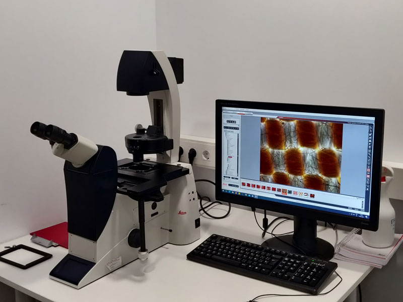
It is an inverted fluorescence microscope, allowing both fixed samples and cell cultures to be viewed.
It has 4 objectives: 10x, 20x, 40x and 63x, the latter being an oil objective.
It has three filters for the visualisation of markers that emit in blue, green and red.
To acquire fluorescence images, this microscope has a camera (Leica DFC450 C) that combines sensitivity and image quality. It also captures bright-field and phase contrast images for microscopy in the life sciences and for clinical and industrial applications.
- Cell distribution studies.
- Cell and tissue morphology studies.
- Cell viability and proliferation studies.
- Research and diagnosis of diseases by immunofluorescence.
ISO 9001
This equipment can be used by users after appropriate training by technicians.
- Priego Villanueva, Sonia
- PTGAS-Ets Investigacio-Microscopia
- Ibañez Gonzalez, Antonio Jose
- PTGAS-Ets Investigacio-Microscopia


