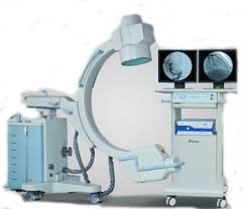

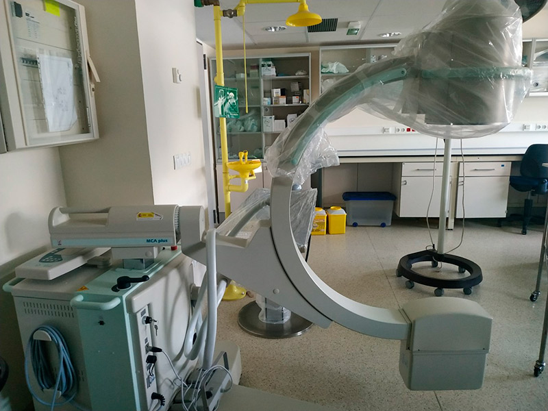
Type: Equipment
Fluoroscopy is the method of real-time X-ray imaging, considered to be a very effective tool for a wide variety of diagnostic examinations and patient interventions. It should not be confused with conventional radiology, which is based on static imaging, commonly known as radiography.
Fluoroscopy is the method of real-time X-ray imaging, considered to be a very effective tool for a wide variety of diagnostic examinations and patient interventions. It should not be confused with conventional radiology, which is based on static imaging, commonly known as radiography.
In fluoroscopy, as in all X-ray imaging, the minimum exposure required for all types of imaging depends on the specific information that needs to be seen or analysed in the image. For this reason, the times required from the image series can be quite long, which affects both the patient and the staff proportionally.
From an occupational health point of view, this method requires the presence of healthcare personnel or staff in the same environment where the radiation is generated, usually a procedure room. This immediately poses a risk of exposure to secondary radiation (scattering), generated by the patient himself or herself each time a fluoroscopic image needs to be produced.
The photons generated within the X-ray tube contain a spectrum of energies. The lower energy photons do not contribute to the image formation and are therefore filtered out using aluminium and copper foil.
The resulting “useful beam” is collimated by leaded apertures to exit the tube housing to the patient, some distance away.
The photons enter the body, undergo attenuation (absorption and scattering to varying degrees by tissues), exit the patient and are detected to form the image required for diagnosis, with appropriate contrast and brightness.
The energy and quantity of X-rays generated in fluoroscopy are mainly controlled by three operating parameters:
- Kilovoltage (kV): Mainly regulates the energy or penetration of the X-ray photons. At the right energy, it provides the necessary contrast for the image.
- Tube current (milliamps, mA): Regulates the number or quantity of photons. An adequate number of photons tends to decrease image noise and provides the required brightness.
- Exposure duration: Depends on how long the exposure is triggered by a foot pedal or hand switch, depending on whether the X-ray beam is continuous or pulsed. Pulsed fluoroscopy emits and cuts the X-ray beam several times per second. A very small amount of the X-rays escapes through the sides of the tube housing and a negligible amount passes through the barrier formed by the metal enclosure of the tube.
The optimal orientation of a fluoroscopy setup is established with the X-ray tube below the patient’s couch and with the detection system above it, usually an image intensifier or a digital flat panel detector.
The Source-Image Distance (SID) is the distance between the focal point and the surface of the image receptor. The SID is fixed on movable C-Arcs at 100 cm. The C-Arc can rotate around its isocentre. The radiation dose increases with the inverse square of the distance (the inverse square law).
The Source-Skin Distance (SSD) is measured from the focal point of the X-ray tube to the entry point of the useful beam into the patient. Therefore, decreasing the SSD increases the risk of radiation-induced skin injury. The minimum SSD, generally allowed in C-Arc, is 30 cm. Many manufacturers provide a spacer in the tube housing, which ensures the 30 cm rule. The spacer must not be removed by the user.
In radiography and fluoroscopy (R and F) installations, the height of the table cannot be changed concerning the X-ray tube. In fixed C-Arc facilities, such as in cardiac catheterisation, special procedure or vascular wards, there are imaging systems that move independently of the tube, providing a variable SID distance in the range of 90 to 120 cm or more. This capability allows us to elevate the imaging detector when space is needed to perform a procedure. In CT fluoroscopy the entire skin surface corresponding to the useful beam width is irradiated uniformly by the rotation of the X-ray tube.
Fluoroscopy is used in medicine primarily for diagnostic purposes, but also some treatment procedures. As a diagnostic test, fluoroscopy allows physicians to study internal organs in real time for signs and symptoms of the disease. As a treatment, fluoroscopy is used to guide surgical procedures that would otherwise be much more invasive.
Among the most common applications are:
In diagnosis
- Digestive system: swallowing studies, oesophagogram, intestinal transit, laparoscopy, barium enema, transcutaneous liver biopsies, enteroclysis, etc.
- Urology: cystography, diagnostic percutaneous nephrostomy, percutaneous excretory urogram.
- Gynaecology: hysterosalpingography.
- Neurology: myelography.
- Cardiology: angiography of vessels of the lower extremities, heart and brain; intracardiac electrophysiological study (EPS), etc.
- Traumatology: arthrography, post-surgical control, etc.
In treatment
- Endoprosthesis placement: oesophageal, biliary, urethral stents, intravascular stents, etc.
- Infiltrations: especially common in intra- and periarticular infiltration of drugs (anaesthetics, corticoids, contrast solutions, etc.).
- Dilatation of vascular, digestive or urological stenosis.
- Guided surgery: biopsies, vertebroplasties, removal of tumours (especially in delicate locations), renal surgery, nephrostomy, placement of pacemakers and cardiac defibrillators, angioplasties, urological surgery (especially in retrograde pyelography).
It should be noted that the exact fluoroscopic procedure may vary considerably depending on the patient’s condition, the structures to be examined, the task to be performed (visualisation or intervention) and the protocols established for each body region. For example, some fluoroscopy-guided interventions may require the use of anaesthesia and patient immobilisation.
ISO 9001
Annual calibration by UV radiation protection service.
Periodic inspections by CSN (Nuclear Safety Centre).
Radioactive source requiring appropriate and legal training.