

Type: Equipment
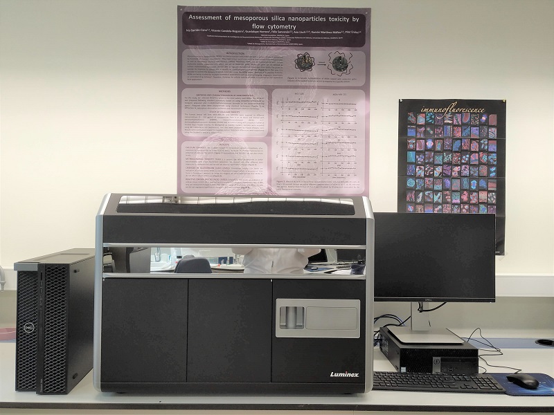
An imaging flow cytometer is a device that combines flow cytometry and microscopy to capture detailed images of cells, allowing a more complete analysis of their morphology and structure.
Imaging cytometry is an innovative and technologically advanced branch of conventional flow cytometry, characterised by new modifications and improvements to existing techniques. Imaging cytometry provides comprehensive information about the morphology or structural features of the cell. This includes a detailed analysis of the size, shape, internal structure and distribution of specific components or markers within the cell. The combination of imaging technologies with flow cytometry therefore represents a breakthrough in cell analysis, providing a complementary source of information to validate fluorescence data and provide a complete characterisation of cell populations. This is a significant advance over conventional flow cytometry, which is largely limited to measuring fluorescence intensity without providing any contextual picture of the cellular structures that produce these signals.
-
Study of cell death/apoptosis/necrosis.
-
Study of reactive oxygen and nitrogen species generation in human and animal models (mouse, rabbit).
-
Study of fluorescent proteins in different human cell lines.
-
Cell phenotype of leukocyte populations, erythrocytes, platelets in human and mouse models.
-
Study of the cellular composition of tumours in human samples as well as in experimental animals.
-
Study of exosomes in cell cultures and plasma samples.
-
Study of cell physiology. Plasma membrane potential, intracellular calcium levels, mitochondrial membrane potential, cell cycle in intact cells and isolated nuclei.
-
Study of bacterial physiology. Cell death, generation of reactive oxygen species, plasma membrane potential.
-
Study of fluorescent molecules bound to certain cell receptors for identification of a specific cell type.
-
Studies of intracellular localisation of transcription factors.
-
Study of phagocytosis.
-
Study of protein co-localisation.
-
Study of transcription factors (cytoplasm-nucleus).
ISO 9001
In order to use this resource, a request must be sent by e-mail or telephone to the Cytometry Service of the UCIM, or through the service platform. The request may also be made in person to the technician.
- Javega Martinez, Beatriz
- PIT-Tecnic/a Sup Pers Tecn Suport
- Escrig Domenech, Aaron
- PTGAS-Etb Investigacio-Biolog.,Quim.,o Sanit.


