Type: Equipment
This platform has various imaging scanners for experiments with small animals (mice and rats). These scanners can also be used to obtain images in solid biological samples.
Image fusion allows the PET/CT/MRI camera to perform both studies simultaneously, providing images that are clearer and contain more information for a more accurate diagnosis. Some examples are:
- anatomical imaging shown by CT/MRI.
- morphological imaging shown by MRI.
- cell function (metabolism) imaging provided by PEDO or IVIS-X5.
Furthermore, imaging agents are needed for some qualitative and quantitative information-gathering techniques.
PET requires radiopharmaceuticals and IVIS needs bioluminescent or fluorescent agents.
Users can also access the corresponding programmes to perform the quantitative analysis for each technique.
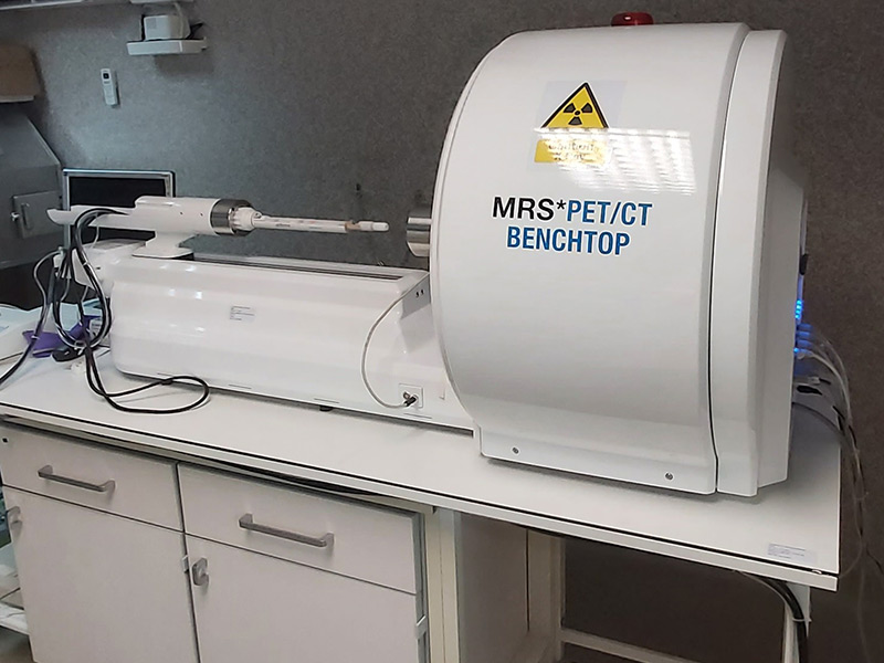
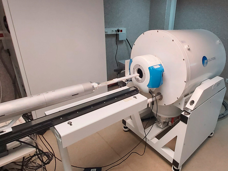
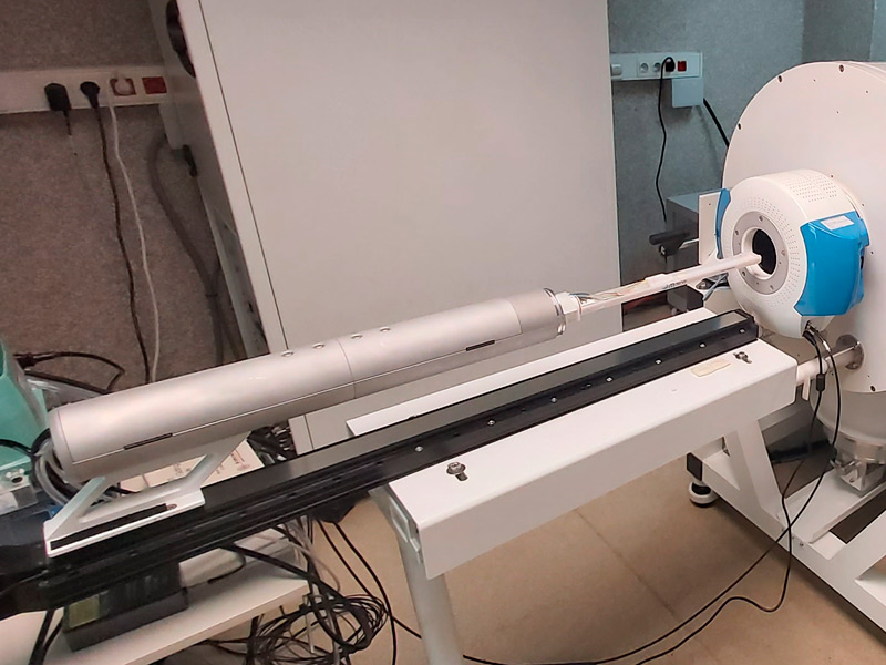
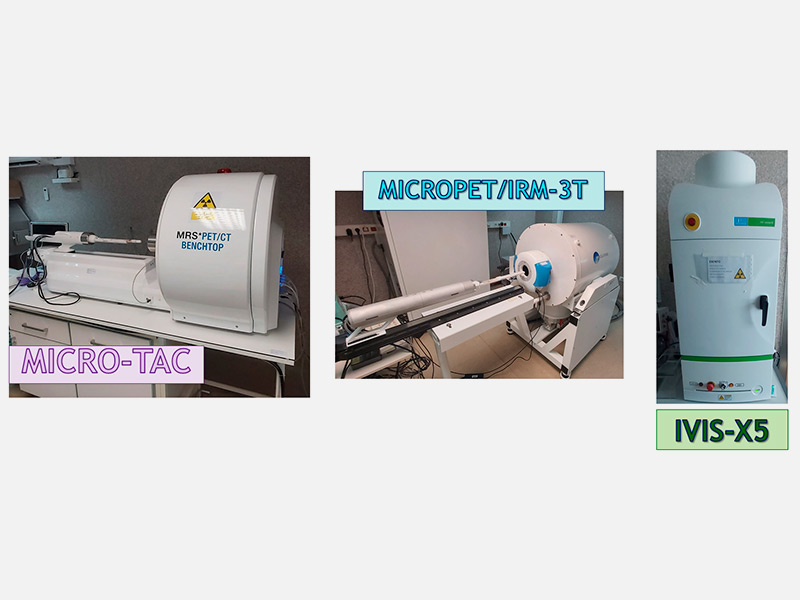
Micro PET/CT/MRI-3T:
This is a hybrid imaging scanner that combines Positron Emission Tomography (PET) and Computed (Axial) Tomography (CT). On the one hand, after injecting radiopharmaceuticals such as 18F-FDG, PET will provide images that give information about cell metabolism. On the other hand, CT is a system of anatomical 2D and 3D imaging of internal organs. This technique uses X-rays to obtain anatomical sections or planes for diagnosis.
MRI scanners are particularly pertinent to obtaining images of non-bone parts or soft tissue. The brain, spinal cord, nerves, as well as muscles, ligaments and tendons are more clearly visible using MRI than using X-rays and CT. For this reason, MRI is often used to image knee and shoulder injuries. The PET scanner can be attached to the MRI scanner, allowing hybrid PET-MRI imaging.
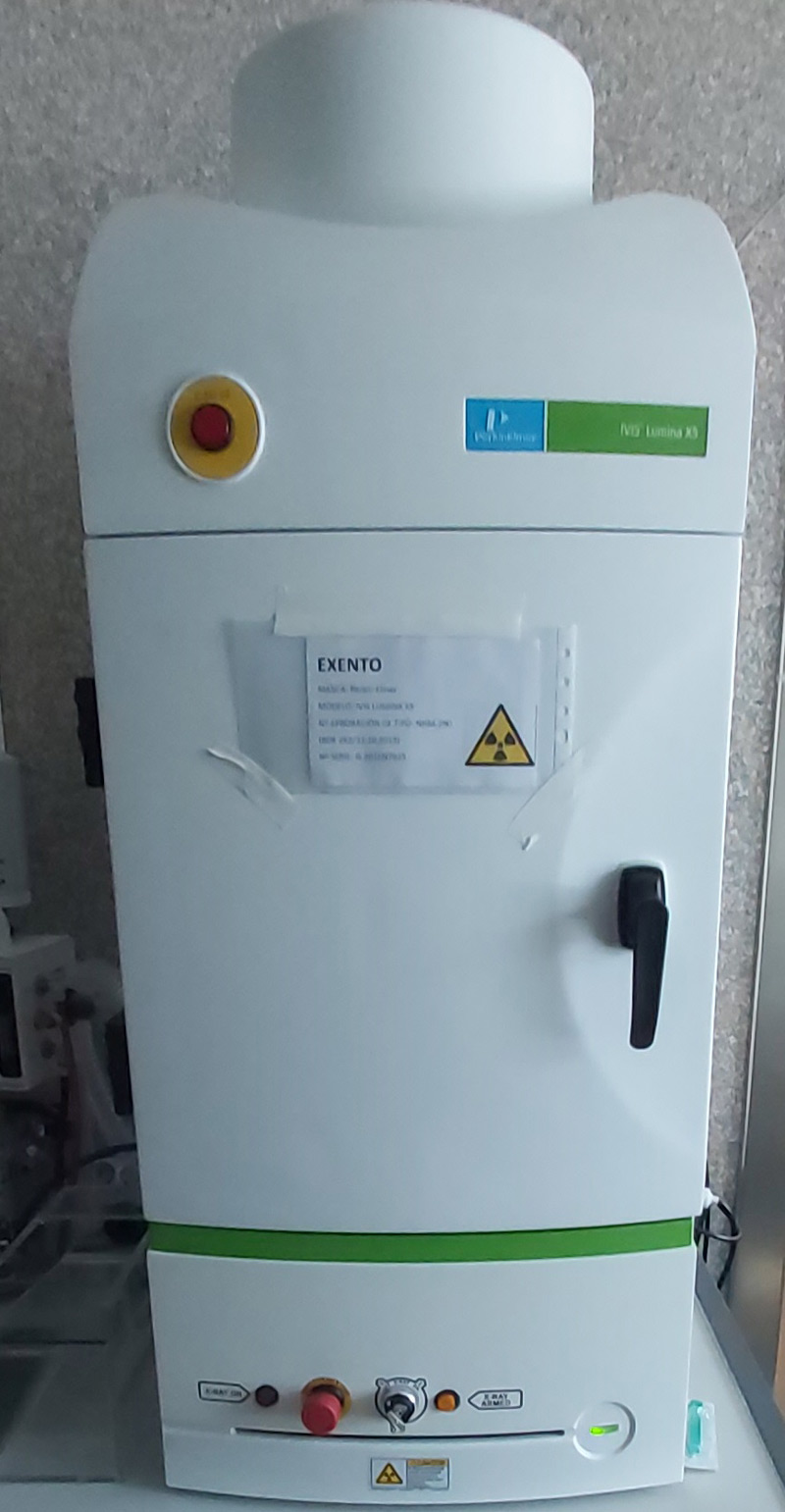
IVIS:
This system makes it possible to obtain and analyse optical images using bioluminescent and/or fluorescent agents. It can also perform 2D imaging using X-rays and Cherenkov imaging.
The PET/CT/MRS-3T system is located at a Unique Scientific and Technical Facility (ICTS) named NANBIOSIS:
This does not add to the cost of our service, but instead gives us access to grants to improve our equipment and, therefore, the service we provide.
Furthermore, being a user of an ICTS means more recognition – it could be said that it is rather prestigious, since there is a selection process. The only requirement is to fill in a form on the NANBIOSIS website, providing an estimate of the number of hours that the service will be required during the semester (or the current year).
There will be another call in June when the number of hours can be readjusted (in case that the actual use is higher than estimated)
This estimation does not imply a commitment to use or pay for the services. The only condition is to mention the Central Service for Experimental Research (SCSIE) in the acknowledgements of the possible works derived from our services by including a sentence, such as: NMR is registered at the U26 facility of ICTS NANBIOSIS at the Universitat de València.
The scanners have a wide range of applications. Studies can be carried out at different levels:
- bone diseases – tumours, fractures, arthritis, osteoporosis, etc.
- cardiovascular diseases – blood vessel disorders, myocardial infarction, etc.
- oncology – all types of tumours
- neurological disorders – dementia, Alzheimer's, Parkinson's, convulsions-epilepsy, etc.
- others – infection or inflammation studies, aneurysm studies, etc.
They can also be used to monitor diseases, study the effectiveness of new medicines, distribute the imaging agent, etc.
- User:
- The user must be informed that the technique makes use of ionising radiation (X-rays). We do not recommend that pregnant individuals attend the study. Nevertheless, the scanners are located in a completely empty room. This means that both the user and the staff are not present when the system is operating
- Animals:
- They may require an adaptation process of 1-5 days if they come from outside of the section, depending on the type of study. Additionally, it is necessary to present a certificate stating the absence of diseases that could contaminate the zone.
- They need to be strictly identified, and the user must provide our staff with a list of the mice with their identification number.
- If a contrast agent is to be used, the animal must be prepared in accordance with the contrast agent technical data sheet.
- In case that it's a new study, the user can request an appointment by email or phone to receive guidance and understand their options.
- Book an appointment on the website:
- New users will need to ask the person in charge to be registered.
- Ezzeddin Ayoub, Mustafa
- PIT-Tecnic/a Sup Uv