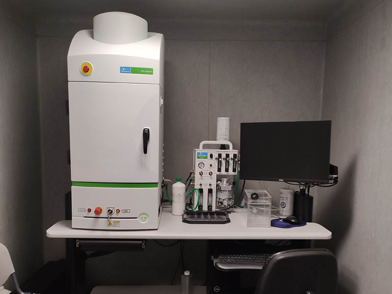

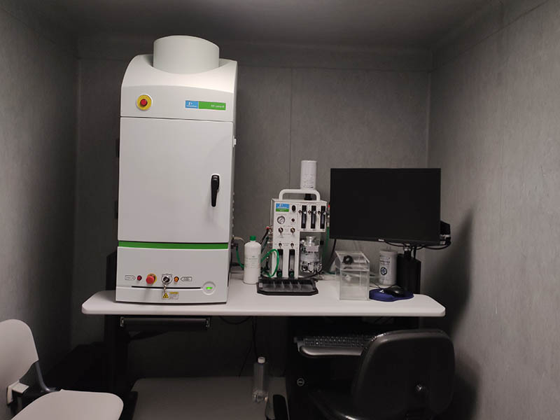
Type: Equipment
The IVIS Lumina X5 combines a high-performance fluorescence and bioluminescence imaging system with a high-resolution 2D X-ray system.
In vivo optical imaging technology allows the non-invasive capture, measurement and analysis of light produced by biological or chemical phenomena inside a living animal, offering spatio-temporal visualisation of biological phenomena. Optical imaging is based on the use of probes such as Luciferase, fluorescent proteins and synthetic fluorophores. Genes coding for Luciferase and fluorescent proteins can be transferred by genetic engineering techniques into cells (e.g. cell lines and pathogens) and animals (transgenic mice and rats) to produce light that can be visualised through living animal tissue using specialised equipment and subsequently analysed using specific software.
- Characteristics:
- High optical and X-Ray performance (allows 5 mice) 20x20cm FOV.
- 2D high resolution, low dose X-ray with optical overlay (anatomical).
- Allows imaging of rats and mice up to 500 - 600g.
- Compute Pure Spectrum (CPS) spectral unmixing.
- Total fluorescence tuning through the Near Infrared (NIR) spectrum.
- Accessories to streamline workflow, data acquisition and analysis.
- Equipped with 26 tunable filters to visualise fluorescent sources emitting from green to near infrared.
- Inside the IVIS Lumina X5:
- The 2.7 x 2.7 cm grade 1 CCD with high quantum efficiency backlighting across the visible to near infrared spectrum.
- Light-tight imaging camera.
- Support for 19 excitation filters and eight emission filters - CPS spectral unmixing.
- LED lamps for photographic imaging.
- Thermal support to maintain optimal body temperature.
- Motor-controlled stage, filter wheels, lens position and f-stop.
- X-ray module:
- Supports small and large rodent models.
- Highly sensitive camera allows fast X-ray image acquisition times of 1-10 seconds reducing radiation exposure.
- Radiation shielded cabinet.
- Exceeds standards set by the US FDA Center for Devices and Radiological Health (21 CFR 1020.40).
- Automated image integration to overlay bioluminescence, fluorescence and photography.
Depending on the type of bioluminescent and/or fluorescent agent it can be used at different levels:
- Inflammatory and infectious processes.
- Cardiological pathologies.
- Pulmonary inflammation.
- Studies of bone development and loss.
- Oncological level: tumour evolution and localisation of metastasis through the marking of different cell lines.
- Other diseases: angiogenesis, arthritis, atherosclerosis...
- Multimodal studies with bioluminescence and fluorescence.
- Preclinical imaging in drug discovery and development.
Certificate ISO 9001
- The user must be informed that the technique uses ionising radiation (X-rays). In the case of users, if they are pregnant, we recommend that they should not be present during the study. However, the equipment is self-armoured in compliance with CSN regulations.
- To reserve the equipment, please arrange a time and day with the section staff by e-mail (see contact) indicating the time, day and number of animals.
- Depending on the study and the bioluminescent/fluorescent agents used, the animal must be prepared before starting the imaging study.