Type: Equipment
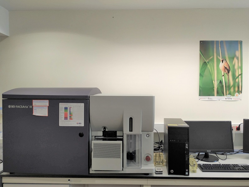
Equipment that determines and quantifies the fluorescence of fluorescent probes attached to different parts of both eukaryotic and prokaryotic cells, organelles, proteins. The emitted fluorescence is collected on different filters limited to a certain wavelength. Fluorescence quantification is associated with the cell parameter being measured, generating quantitative results such as fluorescence intensity, percentage of cell populations, cell count/ML.
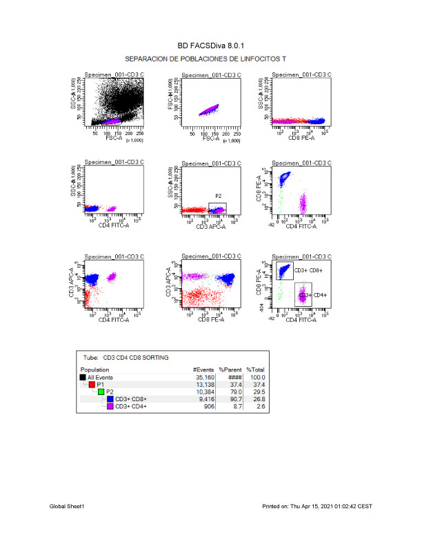
The equipment has a cell separation system allowing the physical separation of a given population with different levels of purity. It allows separation under sterile conditions.
Flow cytometer analyser. The equipment consists of 5 lasers. The configuration of lasers and filters is as follows:
- Blue Laser 488nm contains 2 detectors for Forward Scatter (FSC) and Side Scatter (SSC) cell morphology and 2 filters of 502LP, 530/30 and 655LP 710/50 for fluorescence.
- Violet Laser 405nm contains 5 detectors 735LP 780/60 nm; 630LP 695/40nm; 610LP 660/20nm; 595 LP 616/23nm; 502LP 510/50nm for fluorescence.
- Yellow/Green 561nm laser contains 5 detectors 750LP 780/60 nm; 690LP 710/50nm; 630LP 670/14nm; 600 LP 610/20nm; 585/42 for fluorescence.
- Red Laser 640 nm contains 3 detectors 750LP 780/60 nm; 690LP 730/45nm; 670/30 nm.
- UV 355nm laser contains 2 detectors 710LP 730/45 nm; 660/20nm for fluorescence.
Available with 70, 85, 100 µm nozzle.
Manual sampling. Allows sample collection in different tube formats as well as in 96-well plates.
DIVA 8.0 acquisition software.
- Study of cell death/apoptosis/necrosis.
- Study of reactive oxygen and nitrogen species generation in human and animal models (mouse, rabbit).
- Study of fluorescent proteins in different human cell lines. Separation of populations with a low percentage of positivity.
- Cell phenotyping of leukocyte, erythrocyte and platelet populations in human and mouse models. Separation of different populations.
- Study of cellular composition of tumours and human and mouse tissues. Separation of different populations.
- Study of cell physiology. Plasma membrane potential, intracellular calcium levels, mitochondrial membrane potential, cell cycle in intact cells and isolated nuclei.
- Study of bacterial physiology. Cell death, generation of reactive oxygen species, plasma membrane potential. Library selection in E.coli.
- Study of fluorescent molecules bound to specific cell receptors for identification of a specific cell type.
- Study of specific proteins from mouse neuronal tissue. Cell separation of populations positive for the specific protein.
ISO 9001
Equipment for sample determination. In order to use this equipment, the user must request it by e-mail or by telephone to the Cytometry Service of the UCIM. Samples will always be purchased by the service technician.
- Herrera Martin, Guadalupe
- PIT-Investigacio Escala Tecnica Superior
- Escrig Domenech, Aaron
- PAS-Etb Investigacio


