
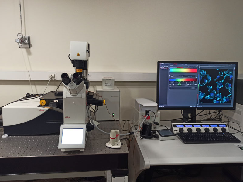
Type: Equipment
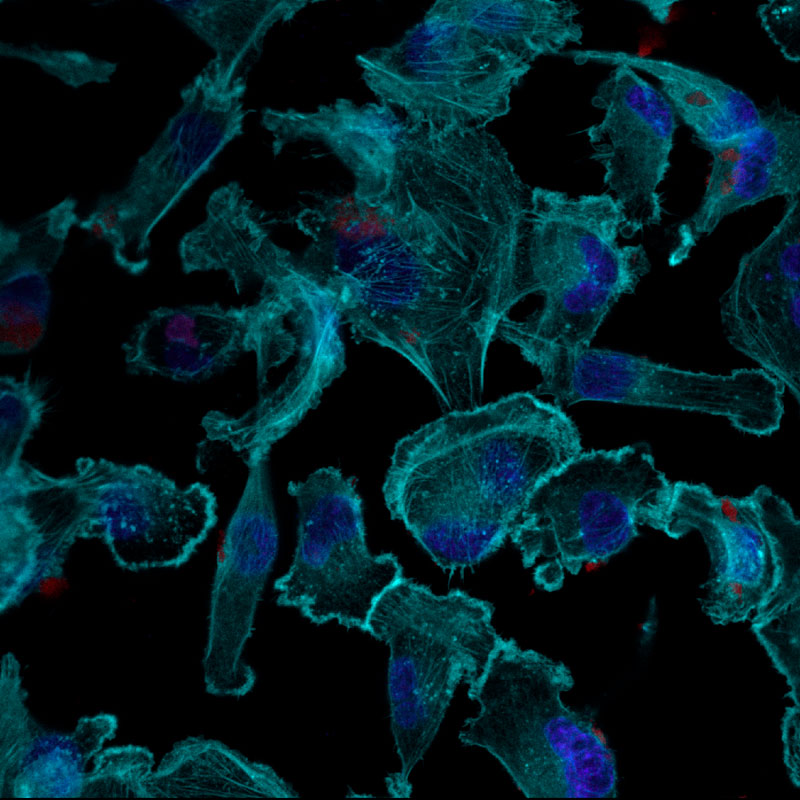
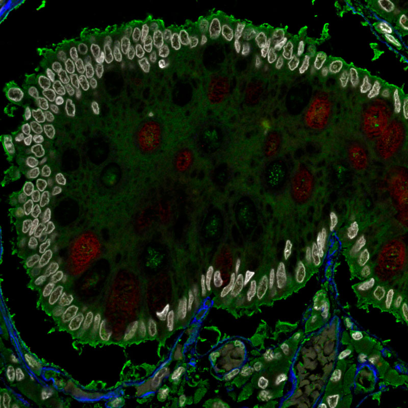
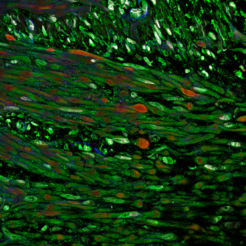
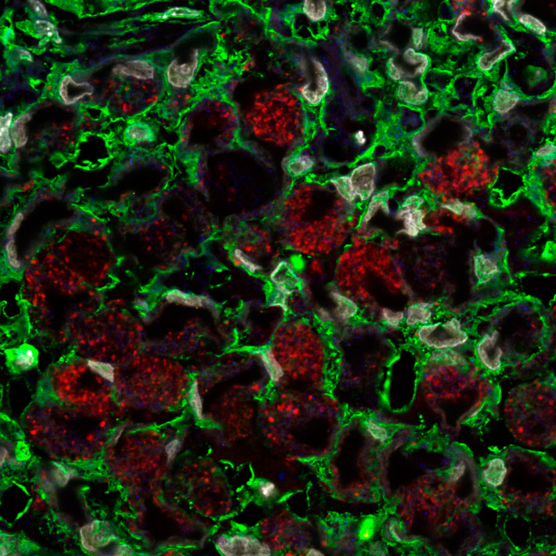
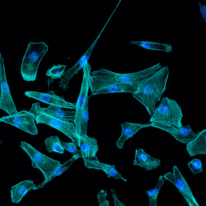
The confocal microscope allows us to visualize images with different fluorescent labelling, therefore obtaining images of great sharpness and quality because the pictures earned are not contaminated by light emitted outside the focal plane. Thanks to this confocal characteristic, we can perform three-dimensional reconstructions from optical sections.
High-end inverted microscope with 5 detectors, one of them for transmitted light.
Thanks to its laser (white laser), it covers almost the entire excitation spectrum.
It incorporates three fluorescence filters to visualise samples that emit in blue, green and red.
As it is inverted, it can be used both to fix samples and to fix live cells. It also includes a stage incubation system to control temperature, humidity and CO2.
It has 5 lenses: 5x, 10x, 20x, 40x(oil) and 63x(oil).
It has a module called Hy-Volution, a system that provides us with high resolution images, showing details of superb quality, improving the axial and lateral resolution of the confocal images obtained.
- Localisation of proteins, organelles, cells and tissue structures using fluorescent probes. tisulares mediante sondas fluorescentes.
- Three-dimensional analysis of biological samples: immunofluorescence in cells, tissues, biofilm.
- Studies of localisation, internalisation and cellular traffic.
- Material studies.
- Analysis of cells in vivo and in real time using markers and/or fluorescent diffusion proteins (GFP) and derivatives.
- Time-lapse (sequences of images obtained over time).
- Viability, proliferation and cell death studies.
ISO 9001
This equipment can be used by users after receiving the appropriate training by the technicians.
- Priego Villanueva, Sonia
- PTGAS-Ets Investigacio-Microscopia
- Ibañez Gonzalez, Antonio Jose
- PTGAS-Ets Investigacio-Microscopia