
A team from the Institute of Corpuscular Physics (IFIC/UV-CSIC) is developing a photon detection and imaging system that will allow visualising, during a medical treatment, the distribution of the radiopharmaceutical in the patient’s body. This technology verifies the final destination of the drug and allows a better estimate of the radiation dose received by both the tumour itself and the vital organs.
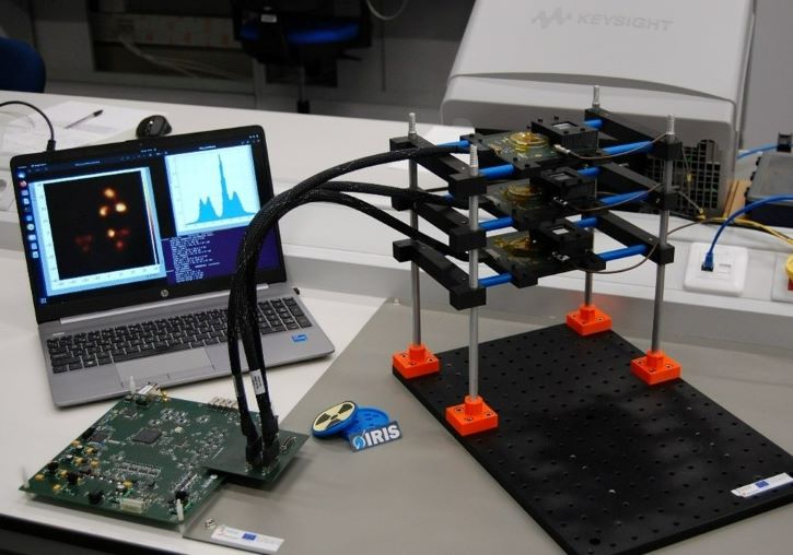
The technology developed by the IFIC’s IRIS (Image Reconstruction, Instrumentation and Simulations for medical applications) group consists of a photon detection and imaging system based on lanthanum bromide crystals coupled to silicon photomultipliers. This method uses two to three sensors in temporal coincidence and, compared to conventional systems, offers higher detection efficiency, very good spatial resolution, and a large field of view.
“We also have a noise reduction method that allows us to work in adverse low-signal scenarios, and we are also using artificial intelligence to improve the image”, says Gabriela Llosá, coordinator of the IRIS group of medical physics at the IFIC, a team specialised in the development of detectors for medical applications, which works in medical imaging and monitoring of hadronic therapy or verification of radiopharmaceutical treatments, in the latter case with the aim of improving the visualisation of its distribution in the body human when administered to the patient.
“To find out the incident energy of the photons in our detector, we have developed analytical imaging models that improve traditional reconstruction algorithms and allow us to obtain four-dimensional images, where the fourth dimension is the energy of the incident gamma rays” adds Jorge Roser, also a researcher at IRIS-IFIC. This technology has previously been used in astroparticle experiments, as well as for the detection of radioactive sources after nuclear accidents aboard drones or robots.
In the medical field, the IRIS group has carried out experiments in collaboration with proton therapy centres such as Quirónsalud (Madrid) to monitor hadronic therapy, and with La Fe Hospital (València) to verify treatment with radiopharmaceuticals. Currently, this technology is in the evaluation and testing phase in relevant environments to increase its TRL – a method for estimating the progress of a technology – with a view to its next commercialisation.
The initiative is part of the VALMONT (INNVA1/2021/37) and VALID (PDC2021-121839-I00) projects, funded by the Valencian Innovation Agency and the State Research Agency, respectively.