
Determination of the cellular fatty acid composition of microbial strains using gas chromatography.
Equipment of the Experimental Aquarium Plant Laboratory.
Nine water filtration and recirculation systems for the preservation of aquatic organisms.
High-throughput platform for High Content Screening image acquisition and high-content analysis. It enables automated acquisition, analysis, and management of fluorescence, brightfield, and digital phase contrast images in multiwell plates, providing a powerful solution for cellular phenotyping and quantitative studies. It supports the study of both live and fixed cells, the characterization and analysis of subcellular compartments, as well as 3D spheroid imaging.
Nebuliser system for respiratory exposure to nanoparticles.
Climate chamber equipped with photoperiod, humidity and temperature control for maintenance and breeding of aquatic invertebrates.
Laboratory equipped with optical material with photographic equipment, growth chamber, specialised bibliography in bryology and computer equipment.
Radiometric calibration laboratory.
Cell Separator equipped with 6 lasers, biosafety cabinet and aerosol extraction. It allows, firstly, the rapid measurement of morphological and functional parameters of a large number of individual cells suspended in liquid medium. For this, cells are initially labelled with fluorochrome-conjugated antibodies, or fluorescent probes that bind to proteins located on the cell surface or inside the cell. The scattered light and fluorescence emitted by the markers as the cells are passed one by one through the different lasers allows simultaneous measurement of structural features and various parameters in each of these cells.
In addition, after analysis, it is possible to separate and collect subpopulations of interest from the sample for further studies.
Experimental Aquarium Plant, Scanning Electron Microscope, Transmission Electron Microscope, Inductively Couple Plasma Mass Spectrometry (ICP-MS), Electrothermal Atomic Absorption Spectrometry (ET-AAS), Sequencing
Climatic chamber with 12 glass aquariums for pathology tests with aquatic organisms.
It contains more than 10,000 samples of parasites (helminths and arthropods) from cetaceans, pinnipeds, sea turtles and fish from the Mediterranean, North Atlantic, Atlantic Ocean, Pacific, Arctic and Antarctic.
A collection of rotifer strains characterised ecologically is available and can be used for basic research, aquaculture centres and toxicity assays.
Collections of seaweed sheets, lichens, lichenicolous fungi and bryophytes included in the VAL herbarium.
Isolated populations from the field or well supplied by reputable collections can be cultivated in our chambers in order to be used in research. Different types of culture techniques are used: individual, mass, semi-continuous and continuous (chemostats).
Storage service for biological material under controlled conditions.
Fully equipped laboratory for sample preparation, acquisition, separation and analysis by flow cytometry.
Genetic analysis with high-performance servers, server-attached computers, bioinformatics software, development of working algorithms.
Life table analysis, monitoring of experimental dynamics, population viability analysis...
The Digital voltmeter Millicell® ERS 3.0 is a devise designed to precisely measure the transepithelial electrical resistance (TEER), the membrane potential in cell cultivations of epithelium and endothelium and the electric the electric potential through the cell monolayers in cultivation inserts. These measures are fundamental to evaluate integrity and confluence of cell monolayers in barrier models, such as the intestine, lung, or the hematoencephalic barrier.
This service provides the group with an aquaria plant, mass spectrometry analysis, electron microscopy and genomics.
Laboratories with gas extraction and laminar flow seeding chamber, equipped with optical and inverted microscopy, centrifuges, spectrophotometer, rotary evaporator, tissue homogeniser, precision scales and other equipment.
Equipment for the analysis of metals and inorganic elements.
A 5-laser analysing cytometer for rapid measurement of morphological and functional parameters of large numbers of single cells suspended in a liquid medium. For this purpose, cells are initially labelled with fluorochrome-conjugated antibodies or fluorescent probes that bind to proteins located on the cell surface or inside the cell. The scattered light and fluorescence emitted by the markers as the cells are passed one by one through the different lasers allows simultaneous measurement of structural features and various parameters in each of these cells.
A three-laser analysing cytometer for rapid measurement of morphological and functional parameters of large numbers of single cells suspended in liquid medium. For this purpose, cells are initially labelled with fluorochrome-conjugated antibodies or fluorescent probes that bind to proteins located on the cell surface or inside the cell. The scattered light and fluorescence emitted by the markers as the cells are passed one by one through the different lasers allows simultaneous measurement of structural features and various parameters in each of these cells.
Qualitative and quantitative analysis of microbial populations by flow cytometry.
Equipment that determines the fluorescence of a substance or material that is capable of absorbing energy in the form of electromagnetic radiation and then emitting part of that energy in the form of electromagnetic radiation of a different wavelength.
Fluorescence inverted microscope that allows for the visualisation and registration of fluorescent markings on cell cultures.
Large climatic chambers where large-capacity microcosm experiments, as well as the cultivation and maintenance of planktonic and macrophyte populations, can be carried out. These cameras allow us to control the light conditions and temperature of the environment and the installation of lamps to obtain radiation of different light spectrum.
Gas chromatographer with single quadrupole mass spectrometry detector, GC-MS, applying volatile compound analysis.
Genotipado de organismos del plancton (SNPs, microsatélites, RFLPs...).
Servers and software for spatially explicit cataloguing and integration of environmental information.
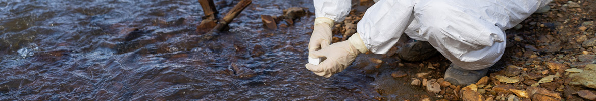
Water and sediment analysis laboratory.
Ability to perform all stages involved in the development of immunoassays for small molecules of the type of natural toxins and agrichemical compounds.
Equipment for the analysis of metals and inorganic elements at trace level.
Air-conditioned chamber equipped with photoperiod, humidity and temperature control for insect maintenance and breeding.
Equipment that quantitatively determines the concentrations of one, several or many substances in a biological (serum, plasma, tissue or urine), food or environmental sample. This is achieved by chromatographic separation of the compounds present in the matrix to be studied, either by liquid chromatography or gas chromatography and their subsequent nebulisation with fragmentation to detect the components according to their molecular weight.
Chamber that allows experimentation with large aquariums and plant cultures.
Research laboratory with a level 2 biosafety chamber.
Laboratory equipped with optical material with photographic equipment, laminar flow cabinet, specialised bibliography on lichens and lichenicolous fungi; TLC equipment (thin layer chromatography) equipped with drying oven, cuvettes and ultraviolet lamp and computer equipment.
Macrophyte live cultures resource.
Technique that allows the lyophilisation of biological material.
Automated cell separation systems based on magnetic separation technology (MACS). It is designed to isolate specific cells in a quick, efficient and reproducible manner from complex samples such as blood, bone marrow or fragmented tissue. It uses detachable magnetic columns and specific reagents to carry out positive or negative cell separations, minimising manual manipulation.
The Massarray platform is responsible for the research study of biological samples through the fine mapping of DNA sequences and the analysis of methylation phenomena. With this equipment, dozens of DNA variants such as SNPs, insertions or deletions can be analysed in hundreds of samples in a short time with a high degree of precision and sensitivity. It is also possible to analyse somatic mutations such as those present in samples of cancerous origin. Finally, the degree of methylation of individual CpGs in short DNA sequences can be quantitatively quantified.
Massive DNA sequencing equipment based on sequencing by synthesis, using nucleotides labelled with fluorophores of different colours, and capturing images of the emitted fluorescence.
Gas accumulation chambers and tubes for experimental measurements, various CO₂ and CH4 measurement systems (absorption spectroscopy, infrared detection -IRGA, semi-conduction).
Microbial characterisation using several approaches.
Collection of pathogenic and non-pathogenic bacteria isolated from different types of samples. Collection of Vibrio vulnificus mutants deficient in virulence genes.
Microbial identification by essential gene sequencing.
The MALDI-TOF MS technique allows obtaining the characteristic protein profile of a microbial strain from the analysis of its ribosomal proteins, mainly.
- Deposit of strains of archaea, bacteria, filamentous fungi and yeasts up to risk group 3* according to the definition of Royal Decree 664/1997 of 12 May 1997.
- Deposit of phages of microbial strains up to risk group 2 according to the definition of Royal Decree 664/1997 of 12 May 1997.
The CECT has a public catalogue that includes more than 8000 strains of bacteria, archaea, filamentous fungi and yeasts. It also has the Mengovirus vMCO and will soon incorporate some phage strains.
Research optical and fluorescence microscopes and stereo microscopes.
Molecular biology tools.
Equipment that performs analysis of metals and inorganic elements.
Standard techniques for observing, describing and measuring animal behaviour.
Ability to design, plan and develop the synthesis of any organic molecule at both laboratory and pilot plant scale.
Laboratory equipped with optical material (binocular magnifying glass and microscope) with digital photographic equipment, specialised bibliography.
Collection of samples and cultures of the different species of Iberian inland waters.
Laboratory equipped with ULT freezer, equipment for molecular biology studies, optical and bibliographic material, computer equipment.
DNA extraction, whole genome sequencing (WGS), assembling, determination of quality parameters, genomic analysis...
A more sensitive and selective piece of equipment, faster in carrying out analysis and a solid service improver for the analysis of trace-level samples (pico-nanograms levels) of a great metabolite number in complex matrices such as biofluids and tissues, in an automated and relatively fast manner, generating a profile by multivariate statistical techniques, etc.
This equipment allows the maintenance of zebrafish or other small size species in controlled conditions.
Real-time PCR is a technique that combines amplification and detection in a single step by correlating the PCR product of each cycle with a fluorescence intensity signal. They consist of a thermal cycler coupled to an optical system, which monitors the signal of the fluorophores used to detect the amplified product. Because the fluorescence of the fluorophores increases as the product is amplified, the amplification and detection processes are combined in a single step.
The UCG has, among other scientific instrumentation, two satellite image reception antennas corresponding to the MSG (Meteosat Second Generation) and NOAA satellites, as well as a reception station for obtaining images from the TERRA, AQUA and NPP satellites (www.uv.es/iplsat/).
The UCG has software licences for satellite image processing.
High-precision spectrofluorometer.
Fluorescence and luminescence diode-array spectrophotometers and multiwell plate readers.
Spectrophotometry (reflectance, transmittance and irradiance) applied to the objective measurement of animal colouration.
Method of sampling aquatic organisms (plankton and charophytes) and their mechanisms of resistance.
The UV Statistics Unit works with experts in the use of computer equipment and specific software. The main software used for data analysis is the free R Software.
The UCG has thermal cameras working in the 8-14 micrometre band for in situ measurement of the earth’s surface temperature.
The UCG has numerous thermal radiometers working in the 8-14 micrometre band for in situ measurement of the earth’s surface temperature.
Table top ultra-centrifuge for small volumes.
UV-Vis spectroscopy equipment based on the process of absorption of ultraviolet-visible radiation (radiation with a wavelength between 160 and 780 nm) by a molecule, which allows us to identify the functional groups present in a molecule.


