
An important migratory route of young neurons in the brains of newborns has been discovered
- Science Park
- February 29th, 2024
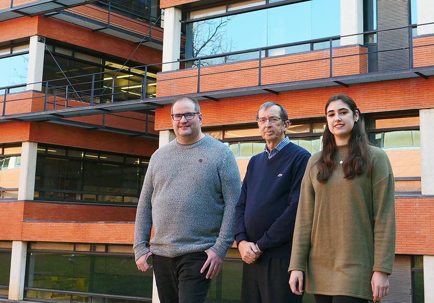
From left to right, Vicente Herranz Pérez, José Manuel García Verdugo, Lucía Torrijos Saiz
A study published in Nature, with notable participation from the University of Valencia, has identified a new migratory route of young neurons in the human infant brain. This migratory pathway extends from the lateral ventricle, where these cells are born, to the entorhinal cortex, an area interconnected with the regions where memory and learning are consolidated. There, these neurons remain dormant in a immature state, awaiting for signals that induce them to mature providing plasticity to the human brain. This migration route begins in the middle of gestation and continues until the child is two or three years old.
Science discovered relatively recently that the production, migration and integration of young neurons in the nervous system did not cease at the time of birth, as previously considered. Instead, certain limited regions maintain this capacity postnatally. The article now published by Nature describes a previously unknown migratory route that begins in the brain of the foetus once gestation is advanced continuing until the child reaches 2-3 years old.
During this period, the cells run their course through the temporal lobe, integrating into the entorhinal cortex a widely interconnected area that participates in the strengthening and stabilisation of memory and learning. There, they remain in an immature state awaiting signals that activate them for their final maturation with the aim of fulfilling a specific mission. This process will help provide the brain with the plasticity necessary to adapt to the experiences that the individual will face throughout life. “Hormonal changes in adolescence, injuties learning new tasks or any type of experience cause internal and external signals that activate neurons and provide plasticity to the brain”, explains Vicente Herranz Pérez (Dep. Cellular Biology, Functional Biology and Physical Anthropology of the University of Valencia) and member of the team that signed the article.
“We were looking for evidence of the existence of young neurons in the human hippocampus and we were surprised by their marked decline between birth and two years of age”, comments José Manuel García Verdugo, professor of Cellular Biology, researcher at the Cavanilles Institute (ICBiBE) of the University of Valencia and co-author of the article. “However, we were able to observe an important flow of immature neurons that converged in the entorhinal cortex. The morphology, the anatomical location, the expression of markers, the distribution and orientation of these cells indicated the presence of an important postnatal neuronal migratory stream in the human brain”, he adds.
This research group has previously participated in the description of other migratory routes in the human brain during early childhood. In this new study, immunostaining, electron microscopy and single-nucleus RNA sequencing techniques have been used; and temporal lobe samples from over 50 donors in late gestation and early childhood ages have been analysed. “This methodology has allowed us to recognise the identity of these cells, their origin and their destiny”, explains Vicente Herranz Pérez. “Interestingly, this population of neurons detected in humans has not been found in species very closely to us, such as macaques, which makes us think that it is a distinctive feature of the human species”, he concludes.
In addition to contributing to the advancement of knowledge in the field of neurogenesis, this finding provides new clues for further research in several lines. On the one hand, it supports the idea of the importance of the plasticity of neuronal circuits. “The late arrival of these young neurons to the areas where memory and learning are consolidated occurs during early childhood, when the child’s interactions with his or her environment are very active”, the article recounts.
On the other hand, the work draws on scientific literature on the early loss of neurons located in this region in Alzheimer’s disease – a fact already accredited by different reports (*1) – to suggest the possible relationship between the alteration in the arrival of this type of cells to the entorhinal cortex and said neurodegenerative pathology. “The entorhinal cortex is one of the first areas affected by Alzheimer’s and this makes us wonder if the neurons that fall by the wayside, or those that do not mature correctly, could be predisposing to the development of this disease”, suggests José Manuel. Garcia Verdugo.
Neuronal migration and maturation, more data for understanding the brain
The publication of this work in Nature coincides in time with another international study, in which the same ICBiBE team participates, which describes in the journal Neuron (Cell Press) the origin of certain immature excitatory neurons present in the amygdala –a brain area related to emotions, fear and anxiety–, this time in both young mice and humans. The work, also focused on the processes of neuronal migration and maturation, resolves that these neurons are not produced by postnatal neurogenesis –as was thought due to the fact that they are still immature cells– but are generated during gestation and they delay their maturation until early adolescence, which means they remain at rest until that stage of development. “During the juvenile age, the majority of the cells in this region undergo structural and physiological maturation, and a subset of them migrate to nearby regions and are activated by various stimuli, which could relate them to plasticity mechanisms”, says Lucía. I. Torrijos Saiz, member of the ICBiBE research team and also co-author of the article in Neuron.
“The relevance of the similarities found between mice and humans lies in the fact that a more accessible animal model has been obtained for the study of this area of the brain, whose alterations could be involved in the appearance of autism spectrum disorders, as already stated. suggested in 2019 (Nature Communications)”, adds the researcher.
Both findings seem to show the growing importance of neuronal migration and maturation processes when advancing our knowledge of the human nervous system. “The future lies in better understanding the dynamics of this type of immature neurons; we need to know how they integrate into pre-existing circuits and what factors cause them to transform into mature cells”, says García Verdugo. “Perhaps, when we are able to identify new populations of immature neurons in other regions, we will have to rethink our current understanding of the mechanisms of plasticity and the functioning of the human brain”, concludes the scientist.
References:
NATURE: Protracted neuronal recruitment in the temporal lobe of young children. Marcos Assis Nascimento, Sean Biagiotti, Vicente Herranz-Pérez, Samara Santiago, Raymund Bueno, Chun J. Ye, Taylor J. Abel, Zhuangzhi Zhang, Juan S. Rubio-Moll, Arnold R. Kriegstein, Zhengang Yang, Jose Manuel Garcia-Verdugo, Eric J. Huang, Arturo Alvarez-Buylla & Shawn F. Sorrells. https://www.nature.com/nature/volumes/626/issues/8001
NEURON: Delayed maturation and migration of excitatory neurons in the juvenile mouse paralaminar amygdala. Pia J. Alderman, David Saxon, Lucía I. Torrijos-Saiz, Malaz Sharief, Chloe E. Page, Jude K. Baroudi, Sean W. Biagiotti, Vladimir A. Butyrkin, Anna Melamed, Chay T. Kuo, Stefano Vicini, Jose M. García-Verdugo, Vicente Herranz-Pérez, Joshua G. Corbinç, Shawn F. Sorrells. https://doi.org/10.1016/j.neuron.2023.11.010
(*1) https://www.cell.com/cell/pdf/S0092-8674(23)00973-X.pdf
https://www.sciencedirect.com/science/article/pii/S0092867423008590?via%3Dihub
More information:
File in: Ciencias de la Vida , Ciencias Médicas